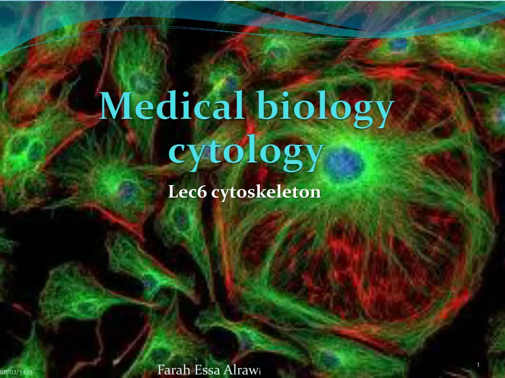
Understanding the Cytoskeleton and Microtubules in Cell Biology
Explore the intricate network of the cytoskeleton and microtubules within cells, essential for structural support, organelle movement, and cell division. Learn how microtubules originate from the centrosome, the dynamic and stable functions of microtubules, and the significance of centrioles in cellular processes.

Download Presentation

Please find below an Image/Link to download the presentation.
The content on the website is provided AS IS for your information and personal use only. It may not be sold, licensed, or shared on other websites without obtaining consent from the author. If you encounter any issues during the download, it is possible that the publisher has removed the file from their server.
You are allowed to download the files provided on this website for personal or commercial use, subject to the condition that they are used lawfully. All files are the property of their respective owners.
The content on the website is provided AS IS for your information and personal use only. It may not be sold, licensed, or shared on other websites without obtaining consent from the author.
E N D
Presentation Transcript
Lec6 cytoskeleton 1 Farah Essa Alrawi 08/03/1439
08/03/1439 2 Farah Essa Alrawi
The Cytoskeleton The Cytoskeleton is a network of tiny protein filaments and tubules that extend throughout the cytoplasm. Functions of cytoskeleton are: Serves the cell s structural framework. 2. Helps maintain a cell s shape . 3. Anchors the organelles . 4. Assists in the movements of organelles and cytoplasmic vesicles. Allows the movement of entire cells. 1. 5. 08/03/1439 3 Farah Essa Alrawi
Microtubules (MTs) Microtubules are found in almost all eukaryotic cell types except red blood cells. They are the largest elements of the cytoskeleton. Microtubules are non branching and rigid hollow tubes of protein that can rapidly disassemble in one location and reassemble in another. 08/03/1439 4 Farah Essa Alrawi
08/03/1439 5 Farah Essa Alrawi
. All microtubules originate from the microtubule- organizing center (MTOC) called centrosome, has gamma tubulin ( ). Centrosome is an area of the cytoplasm located near the nucleus Microtubules are elongated polymeric structures composed of equal parts of tubulinand tubulin. Microtubules grow from tubulin rings within the MTOC that serve as nucleation sites for each microtubule. The length of microtubules changes dynamically as tubulindimers are added or removed in a process of dynamic instability 08/03/1439 6 Farah Essa Alrawi
In the centrosome, the tubulin subunits polymerize and from two types of microtubules: 1. Dynamic microtubules are continuous assembly and disassembly (reshaping of cell) determine cell shape and function in intracellular movement of organelles and secretory granules and form spindles that guide the movement of chromosomes during cell division or mitosis 2. Stable microtubules form walls of centrioles, cilia and flagella. They are responsible for the beating movements 08/03/1439 7 Farah Essa Alrawi
Stable microtubules 1. Centrioles. 2. cilia . 3. flagella. 08/03/1439 8 Farah Essa Alrawi
Centrioles Centrioles are non membranous organelles. Small cylindrical structures composed of highly organized microtubules located within centrosome, perpendicular to each other. Each centrioleconsists of nine evenly spaced clusters of three microtubules arranged in a circle. The microtubules have longitudinal orientation and are parallel to each other. 08/03/1439 9 Farah Essa Alrawi
08/03/1439 10 Farah Essa Alrawi
08/03/1439 11 Farah Essa Alrawi
Before mitosis, the centrioles in the centrosome replicate and form two pairs. During mitosis, each pair moves to the opposite poles of the cell, become microtubuleorganizing centers for mitotic spindles that control the distribution of chromosomes daughter cells. where they to the 08/03/1439 12 Farah Essa Alrawi
Several biologists to study details of microtubule dynamics are also widely used in cancer chemotherapy to block activity of the mitotic spindle in rapidly growing neoplastic cells. Such drugs include vinblastine and vincristine. Vincristine works partly the tubulin protein, stopping the cell from separating its chromosomes during the metaphase the cell then undergoes apoptosis. The vincristine molecule inhibits leukocyte maturation inhibitory compounds used by cell by binding to production and 08/03/1439 13 Farah Essa Alrawi
08/03/1439 14 Farah Essa Alrawi
08/03/1439 15 Farah Essa Alrawi
Cilia Cilia (sing., cilium) are involved in movement. Motile structure use to move something like the ciliated cells that line our respiratory tract sweep debris trapped within mucus back up the throat. This helps keep the lungs clean by rhythmic beating. Similarly, ciliated cells move an egg along the oviduct, where it will be fertilized by a flagellated sperm cell. 08/03/1439 16 Farah Essa Alrawi
Origin of cilia from centrioles, each centrioles give only one cilium so ciliated cells have many centioles embedded in cytoplasm under cell membrane called basal body. Basal bodies associated structures firmly anchor cilia in the apical cell cytoplasm 08/03/1439 17 Farah Essa Alrawi
08/03/1439 18 Farah Essa Alrawi
08/03/1439 19 Farah Essa Alrawi
Cilia have another function; act as receptor in special cells (rods and cones cells of the eyes retina). 08/03/1439 20 Farah Essa Alrawi
Flagella Flagella (sing. Flagellum) is motile projection use to move cell itself, like tail of sperm. Have an inner core of microtubules within a covering of plasma membrane. Flagellum is the same structure of cilium but always single and extremely longer. 08/03/1439 21 Farah Essa Alrawi
08/03/1439 22 Farah Essa Alrawi
08/03/1439 23 Farah Essa Alrawi
Microtubules essential cellular functions Intracellular vesicular transport (e.g., movement of secretory vesicles, endosomes, and lysosomes). 2. Movementof cilia and flagella. 3. Attachment of chromosomes to the mitotic spindle and their movementduring mitosis and meiosis. 4. Cell elongation and movement (migration). 5. Maintenanceof cell shape. 1. 08/03/1439 24 Farah Essa Alrawi
Some individuals have an inherited genetic defect that leads to malformed microtubules in cilia and flagella. Called immotile cilia syndrome These individuals suffer from recurrent and severe respiratory infections. The ciliated cells lining respiratory passages fail to keep their lungs clean. (chronic respiratory infections) They are also unable naturally due to the lack of ciliary action to move the egg in a female or the lack of flagella action by (Immotile sperm). to reproduce sperm in a male. 25
Filaments There are three types of filaments: 1. Microfilaments. 2. Intermediated filaments . 3. Thick filaments. 08/03/1439 26 Farah Essa Alrawi
Microfilament (thin filament or actin filaments) Microfilaments are the thinnest structures of the cytoskeleton usually occur in bundles or other groupings. They are composed of the protein actin and are most prevalent on the peripheral regions of the cell membrane. Structure of actin protien is fine strands of globular actin (G-actin). 08/03/1439 27 Farah Essa Alrawi
Function of micro filaments: These structural proteins shape the cells. 2. involved in cell movementand movement of the cytoplasmicorganelles. 3. The microfilaments are distributed throughout the cells and are used as anchors at cell junctions. 4. The actin microfilaments also form the structural core of microvilli (non motile cellular membrane protrusions that increase the surface area for diffusion and minimize any increase in volume such as the epithelial cells of small intestines) and the terminal web just inferior to the plasma membrane. 1. 08/03/1439 28 Farah Essa Alrawi
Intermediate filaments as their name implies, are intermediate in size between microtubules and actin filaments. Several cytoskeletal proteins that form the intermediate filaments have been identified and localized. 08/03/1439 29 Farah Essa Alrawi
Their structure and function differ according to the type of cell. Keratain filaments. In skin cells, these filaments terminate at cell junctions, where they stabilize the shape of the cell and their attachments to adjacent cells. Vimentin filaments are found in many mesenchymal cells. Desmin filaments are found in both smooth and striated muscles. Neurofilament proteins are found in the nerve cells and their processes. Glial filaments are found in astrocyticglial cells of the nervous system. Lamin intermediate filaments are found on the inner layer of the nuclear membrane. 08/03/1439 30 Farah Essa Alrawi
The intermediate filament in tumors can often reveal the cellular origin of the tumor, information important for diagnosis and treatment of the cancer. Identification of intermediate filament proteins by means of immunocytochemical routine procedure. One example is the use of Glial Fibrillary Acidic Proteins (GFAP) to identify astrocytomas, the most common type of brain tumor. presence of a specific type of methods is a 08/03/1439 31 Farah Essa Alrawi
Thick filament The thick filaments in muscle tissues are the actin filaments fill the cells and associated with myosin proteins to induce muscle contractions. 08/03/1439 32 Farah Essa Alrawi
08/03/1439 33 Farah Essa Alrawi


")