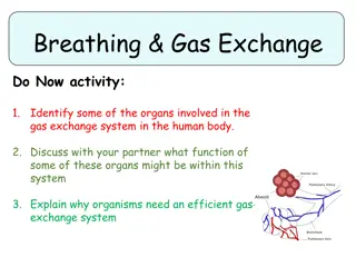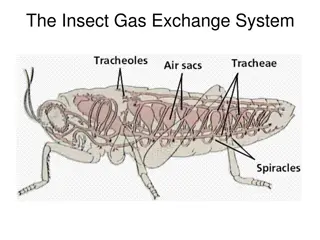
Understanding the Gas Exchange System in Organisms
Learn about the gas exchange system in organisms, including how they take in gases from the environment and release gases, the role of the gas exchange surface in the lungs, the structures involved in gas exchange, and the importance of cartilage, ciliated epithelium, goblet cells, smooth muscle cells, and elastic fibers in the process.

Download Presentation

Please find below an Image/Link to download the presentation.
The content on the website is provided AS IS for your information and personal use only. It may not be sold, licensed, or shared on other websites without obtaining consent from the author. If you encounter any issues during the download, it is possible that the publisher has removed the file from their server.
You are allowed to download the files provided on this website for personal or commercial use, subject to the condition that they are used lawfully. All files are the property of their respective owners.
The content on the website is provided AS IS for your information and personal use only. It may not be sold, licensed, or shared on other websites without obtaining consent from the author.
E N D
Presentation Transcript
All organisms take in gases from their environment and release gases to the environment. Animals take in O2for aerobic respiration and release CO2. Plants also respire, but during daylight hours they photosynthesise at a greater rate than they respire, and so take in CO2and release O2.
The body surface across which gases diffuse into and out of the body is called the Gas Exchange Surface. In mammals, including humans, the gas exchange surface is the surface of the alveoli in the lungs.
The gas exchange surface in the lungs is extensive, very thin, well supplied with blood well ventilated. The trachea and bronchi provide little resistance to the movement of air to and from the alveoli.
Cartilage in the walls of the trachea and bronchi provides support and prevents the tubes collapsing when the air pressure inside them is low. Cillated epithelium is found lining the trachea, bronchi and some bronchioles. It is a single layer of cells whose outer surfaces are covered with many thin extensions (cilia) which are able to move. They sweep mucus upwards towards the mouth, helping to prevent dust particles and bacteria reaching the lungs.
Goblet cells are also found in the ciliated epithelium. They secrete mucus, which traps dust particles and bacteria.
smooth muscle cells are found in the walls of the trachea, bronchi and bronchioles. This type of muscle can contract slowly but for long periods without tiring. When it contracts, it reduces the diameter of the tubes. During exercise it relaxes, widening the tubes so more air can reach the lungs.
Elasticc fibres are found in the walls of all tubes and between the alveoli. When breathing in, these fibres stretch to allow the alveoli and airways to expand. When breathing out, they recoil, helping to reduce the volume of alveoli and expel air out of the lungs.
GAS EXCHANGE SYSTEM The air inside an alveolus contains a higher concentration of O2, and a lower concentration of CO2, than the blood in the capillaries. This blood has been brought to the lungs in the pulmonary artery, which carries deoxygenated blood from the heart. O2therefore diffuses from the alveolus into the blood capillary, through the thin walls of the alveolus and the capillary. CO2diffuses from the capillary into alveolus
The diffusion gradients for these gases are maintained The diffusion gradients for these gases are maintained by by: breathing movements breathing movements, which draw air from outside the body into the lungs, and then push it out again; this maintains a relatively high concentration of O2and low concentration of CO2in the alveoli; blood flow past the alveolus blood flow past the alveolus, which brings deoxygenated blood and carries away oxygenated blood.
Tidal volume and vital capacity.. To be cntd agn The volume of air that is moved into or out of the lungs during one breath is called the tidal volume. It is generally about 0,5 dm3. The maximum amount of air that can be moved in or out during the deepest possible breath is called the vital capacity. It is generally between 3 dm3and 5 dm3.
EIC muscles and of Diaphragm Air moves by mass flow into and out of the lungs during breathing. This is caused by the contraction and relaxation of external intercostal muscles and muscles in the diaphragm. When these contract, they increase the volume of the thoracic cavity and draw air down through the trachea and into the bronchi and bronchioles. When they relax, the thoracic volume decreases and air flows out, down a pressure gradient.
The smoke from cigarettes contains several substances that affect the gas exchange system and the cardiovascular system. These include: tar , nicotine, CO Tar : tar, a mixture of substances including various chemicals that act as carcinogens.
Nicotine, : an addictive substance that affects the nervous system by binding to receptors on neurones (nerve cells) in the brain and other parts of the body. It increases the release of a neurotransmitter called dopamine in the brain, which gives feelings of pleasure. It increases the release of adrenaline into the blood, which in turn increases breathing rate and heart rate.
CO + Hb =CarboxyHb Carbon monoxide diffuses across the walls of the alveoli and into the blood in the lungs. CO + Hb = CarboxyHb ----- reduces oxygen supply when diffuses into RBCs This means that haemoglobin does not become fully oxygenated.
Effects of smoking on the Gas Exchange System Chronic obstructive pulmonary disease (COPD) COPD takes 2 main forms This is a condition in which a person has Chronic bronchitis and Emphysema. It can be extremely disabling.
Chronic bronchitis symptoms : long term cough and phlegm(mucus) 1.components of cigarette smoke, including tar, cause goblet cells to increase mucus production . 2. This causes mucus to build up, which may partially block alveoli 3. This makes gas exchange more difficult, as the diffusion distance between the air in the alveoli and the blood in the capillaries is greater.
The mucus becomes infected with bacteria, causing bronchitis. Smokers often have chronic (long-lasting) bronchitis.(inflammation of the lining of bronchial tubes ) The mucus stimulates persistent coughing, which can damage the tissues in the walls of the airways, making them stiffer and the airways narrower.
Emphysema is a chronic lung disease. Emphysema is characterized by loss of elasticity of the alveoli - destruction of structures supporting the alveoli; - destruction of lung tissue with time
Lungs contain large number of macrophages, from blood circulation. These phagocytes release hydrolytic enzymes(elastase) which break elastic fibres(elastin) of the alveolar walls. Loss of elastin makes it difficult to move air out of lungs
Air remains in the lungs , its not refreshed during ventilation and fails to recoil There is reduced surface area for gas exchange Oxygenation is reduced in emphysema , with rapid breathing rate .
. A person with emphysema has shortness of breath, meaning they struggle to breathe as deeply as they need to, especially when exercising
As emphysema progresses, blood vessels in the lungs become more resistant to the flow of blood. To compensate for increased resistance: BP in pulmonary artery increases and overtime the Rt side of heart enlarges .
As lung function deteriorates , wheezing occurs , breathlessness worsens. People with emphysema often need a continous supply of oxygen to stay alive They cannot get out of bed .
Symptoms of breathlessness becomes troublesome when about half of the lungs are destroyed . If smoking is given up at an early age , lung function can improve .
LUNG CANCER Various components of tar can cause changes in the DNA in body cells, including the genes that control cell division, which can cause cancer. These substances are therefore carcinogens. Cancers caused by cigarette smoke are most likely to form in the lungs but may form anywhere in the gas exchange system, and also in other parts of the body.
A single mutation is responsible for triggering lung cancer, Accumulation of mutations with time causes a group of cells to divide by mitosis repeatedly forming an irregular mass of cells tumour . As cancer develops , it spreads through the bronchial epithelium , and enters the lymphatic tissues in lungs . .
Sometimes tumour cells break away and are carried to other parts of body , forming a secondary tumour (metastasis) Referred as malignant tumour.
Symptoms of lung cancer : coughing of blood ,chest pain, difficult to breathe , fatigue and weight loss. Unchecked , cancerous cells take over the body , leading to malfunction and death
Location of tumours by one of the methods Bronchoscopy using an endoscope to allow direct view of lining of bronchi Chest X ray CT scan
Treatment Include surgery , radiotherapy, chemotherapy It is also dependant on the type of lung cancer and metastasis.
Effects of smoking on CVS The nicotine and CO in tobacco smoke increase the risk of coronary diseases and stroke. Nicotine is the drug in tobacco. nicotine is absorbed very readily by the blood and travels to the brain within a few seconds. It stimulates the nervous system to reduce the diameter of the arterioles To release the hormone adrenaline from the adrenal glands.
As a result, heart rate and blood pressure increase There is a decrease in blood supply to the extremities of the body, such as hands and feet, reducing their supply of oxygen. Nicotine also increases the risk of blood clotting
It stimulates nerve endings in the brain to release the neurotransmitter substance dopamine, Dopamine is associated with reinforcing pleasurable experiences. This makes it very hard to give up smoking.
CO + Hb =CarboxyHb Carbon monoxide diffuses across the walls of the alveoli and into the blood in the lungs. CO + Hb = CarboxyHb ----- reduces oxygen supply when diffuses into RBCs This means that haemoglobin does not become fully oxygenated.
Less oxygen is supplied to the heart muscle, putting a strain on it especially when the heart rate increases during exercise. Carbon monoxide also damage the lining of the arteries.
Damage to the walls of the arteries leads to build up of fatty tissue and reduction of blood flow. Coronary heart disease and stroke are the results .