Understanding the Lacrimal Apparatus and Tear Formation Process
Learn about the lacrimal apparatus, including the lacrimal gland, conjunctival sac, and nasolacrimal duct, which are essential for tear formation and transport in the eye. Explore the functions of the lacrimal glands, accessory glands, and their role in maintaining eye health. Discover the structures involved in tear production, such as the palpebral part of the conjunctiva and the lacrimal canaliculi.

Download Presentation

Please find below an Image/Link to download the presentation.
The content on the website is provided AS IS for your information and personal use only. It may not be sold, licensed, or shared on other websites without obtaining consent from the author. If you encounter any issues during the download, it is possible that the publisher has removed the file from their server.
You are allowed to download the files provided on this website for personal or commercial use, subject to the condition that they are used lawfully. All files are the property of their respective owners.
The content on the website is provided AS IS for your information and personal use only. It may not be sold, licensed, or shared on other websites without obtaining consent from the author.
E N D
Presentation Transcript
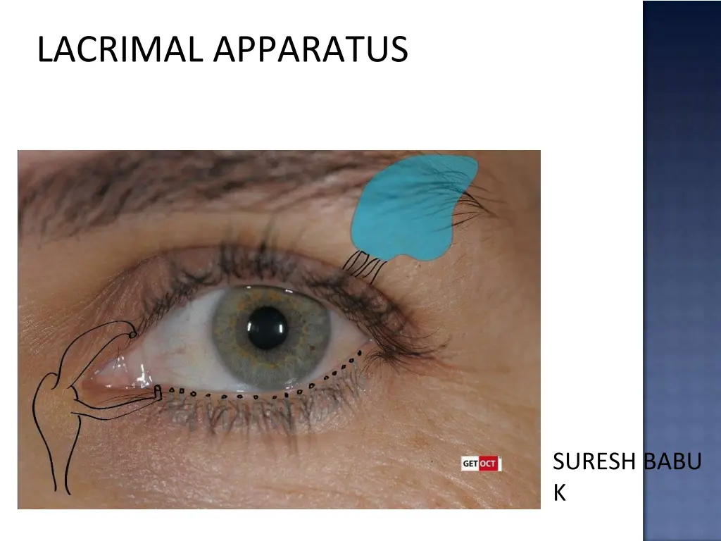
LACRIMAL APPARATUS SURESH BABU K
It is concerned with the tear formation & transport. Lacrimal passage includes : Lacrimal gland Conjunctival sac Lacrimal puncta Nasolacrimal duct Lacrimal sac Lacrimal canaliculi
Conjunctiva = the mucous membrane of the eye. A. Palpebral part = forms a crescentric fold or Plica semilunaris. B. Bulbar( ocular ) = thin, transparent, slightly avascular over the sclera. C. Conjunctival sac = with lids closed, a space between the eyeball and eyelids , lined by conjunctiva. Lacrimal glands = secretes lacrimal fluids Lacrimal ducts = conveys fluids from glands to the conjunctiva sac. Lacrimal canaliculi = connection between lacrimal punctum
It includes lacrimalgland, accessory glands Lacrimal gland is above & anterolateral toglobe. Secretes tears into superiorfornix. Tears moisten & lubricates the : cornea , conjunctiva. It contributes 43D of 50D of refractive power of eye.
It consists of Large OrbitalPart Smaller Palpebral Part Lateral expansion of levator separates the parts
Paired almond-shapedglands. It is present in a fossa on the anterolateral area of orbit It has 2 surfaces, 2 borders, 2extremities Superiorsurface Frontalbone Inferiorsurface Levator palpebrae superioris & lateral rectus
Anteriorborder Septum orbitale Posterior border Contact with orbital fat , level with posterior pole. Lateral extremity Rest on lateral rectus Medial extremity On levator
1/3rdsize of orbitalpart Superior fornix , seen on lideversion. It is situated upon the course ofducts Related to levator superiorly, inferiorly to superior fornix Posteriorly it continues with orbitalpart.
Are small, compound, branched, tubularglands Located in the middle of lid (Wolfring glands) or superior & inferior fornices (Krauseglands). Ectopic portions of lacrimal glandtissue.
It is with connective tissue coat and excretory duct. The excretory duct splits & ducts, connected to secretory glandular epithelia. form intralobular Secretory epithelia have elongatedtubules. True acini are absent.
Artery supply : Lacrimal artery , branch of ophthalmic artery. Venous drainages : Ophthalmic Vein. Lymphatic drainage : Joins that of conjunctiva & drain into the preauricular lymph nodes.
Sensory nerve supply : lacrimal nerve , branch of ophthalmic division of Vth nerve Sympathetic nerve supply : carotid plexus Secretomotor fibers : superior salivary nucleus
A small, round or oval orifice on the elevation, the papilla lacrimalis. At medial end of lid margin at the junction of its ciliated and non-ciliatedparts. Upper punctum medial to lower, from the medial canthus being 6 and 6.5mm. The upper punctum opens inferoposteriorly, the lower superoposteriorly.
First vertical and then horizontal Vertical part is 2 mm & turns medially at right-angle to become horizontal 8 mm At angle - dilatation or ampulla. The canaliculi pierce the fascia (i.e. the periorbita covering the lacrimal sac) separately, Uniting to enter lacrimalsac. Stratifiedsquamous epithelium elastic tissue. supported by
Lacrimal fossa, formed by frontal processof maxilla . lacrimal boneand The sac, closed above and open below, is continuous with the nasolacrimalduct. The sac is enclosed by a periorbita, splits &form the lacrimal fascia .
Relations Medial : periorbita and bone, arc of ethmoid sinuses. Lateral : skin, orbicularis oculi, and lacrimal fascia. Anterior: medial palpebral ligamentand angular vein. Posterior : lacrimal fascia andmuscle
The nasolacrimal duct, continuation of lacrimal sac to the inferior meatus. 15 mm. It lies in a canal formed by the maxilla, lacrimal bone and lacrimal processof inferior concha. It descends posterolaterally, a surface indication a line from medial canthus to first uppermolar.
The valves They are folds of mucous membrane with no valvularfunction. The most constant is the 'valve' of Hasner at the lower end. It prevents sudden blast of air (when blowing the nose) from entenng the lacrimal sac.
Structure Double-layeredEpithelium The superficial layer composed of columnar cells, the deeper cells beingflatter. The membranous wall of the sac is of fibroelastic tissue, the elastic element being continued around thecanaliculi.
Around the nasolacrimal duct is plexus of vessels, forming erectile tissue like that on the inferior concha. Engorgement of these vessel obstruct theduct. The course of the lacrimal sac and duct can be demonstrated bydacryocystography
Vessels Artery supply : palpebral branches of the ophthalmic, angular and infraorbital arteries and nasal branch of the sphenopalatine. Venous drainages : Angular and infraorbital vessels above, below into the nasal veins Lymphatic drainage: submandibular and deep cervical nodes. Nerves Infratrochlear and anterior superior alveolar nerves.
Tears are lost from the conjunctiva sac by absorption, evaporation, and nasolacrimal system. This is related to the size of the palpebral aperture, the blink rate, ambient temperature and humidity.
Tears flow the upper and lower marginal strips lower canaliculi (capillarity+suction) Eyesclose Pretarsal orbicularis oculi compresses the ampullae+ shortens and compresses canaliculi+puncta medially. Lacrimal part of the orbicularis oculi, contracts compresses the sac,(positive pressure) tears nasolacrimal duct nose. Eyesopen Muscles relax canaliculi and sac expand(negative pressure)+capillarity= tears into sac. upperand