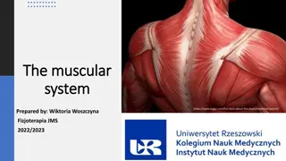
Understanding the Muscular System - Myology Overview
Muscular system encompasses various types of muscle tissue enabling movement and stability in the body. Learn about skeletal muscles, muscle organization, and accessory structures like synovial membranes and fasciae.

Download Presentation

Please find below an Image/Link to download the presentation.
The content on the website is provided AS IS for your information and personal use only. It may not be sold, licensed, or shared on other websites without obtaining consent from the author.If you encounter any issues during the download, it is possible that the publisher has removed the file from their server.
You are allowed to download the files provided on this website for personal or commercial use, subject to the condition that they are used lawfully. All files are the property of their respective owners.
The content on the website is provided AS IS for your information and personal use only. It may not be sold, licensed, or shared on other websites without obtaining consent from the author.
E N D
Presentation Transcript
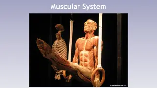
THE MUSCULAR SYSTEM THE MUSCULAR SYSTEM MYOLOGY MYOLOGY The muscular system can be defined as all forms of tissue in the body that can contract to perform movement or similar functions. Muscular tissue is of three kinds: (1) Striated (2) non-striated, or smooth; and (3) cardiac. The striated muscles, being for the most part directly or indirectly connected with the skeleton, are often termed skeletal or somatic, it cover the greater part of the skeleton and red in color. Skeletal Muscles vary greatly in form, and may be classified as (a) Long ;(b) short (c) flat; {d) ring-like or orbicular. Long muscles are present chiefly in the limbs, while the flat or broad muscles occur principally in the trunk, where they assist in forming the walls of the body cavities. The ring-like or orbicular muscles circumscribe orifices which they close, and are hence termed sphincters.
Most movements of the animal body and its parts are caused by muscular contraction. Muscle is also used to prevent movement, stabilizing to prevent their collapse under a load and main taining continence of bladder and bowel. A subsidiary function of the skeletal muscles is to generate shivering , involuntary tremors initiated by exposure to cold. limb
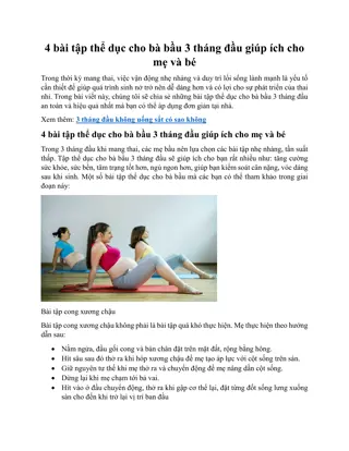
Organization of skeletal muscle : skeletal muscle is butchers meat and accounts for about half a weight of an animals carcass . Each individual muscle is composed of many cells held together by connective tissue . The whole muscle is covered by a dense connective tissue called epimysium , bellow this , a looser layer , the perimysium , covers the small bundles into which the fibers are grouped . Finally , each fiber is provided with its own delicate covering , the endomysium . Attachments: Skeletal muscle is attached to bones directly or by the use of tendons , through the perimysium of the muscle that fuse with the periosteum or perichondrium structures called aponeuroses. In certain positions, especially where tendons play over joints or are subjected to great pressure, sesamoid bones like patella develop in the original tendon tissue. , or intermediary fibrous
Figure 126 Transection of a skeletal muscle; the fibrous tissue has been emphasized. 1, Epimysium; 2, perimysium; 3, endomysium.
The accessory structures connected with the muscles are the synovial membranes and the fasciae. The synovial membranes are arranged in two principal forms: (a) Bursal; (b) vaginal. A bursa (Bursa mucosa) is a simple sac interposed between the tendon or muscle and some deeper seated structure most commonly a bony prominence. A vagina tendinis or tendon sheath is the synovial sac that folded around the tendon more or less completely. The two layers are continuous along a fold termed the mesotendon. The articular synovial membranes in some places form extra-articular pouches, which facilitate the play of tendons. The fasciae are sheets of connective tissue. Two layers may usually be recognized. The superficial fascia is composed of loose connective tissue which may contain more or less fat and is subcutaneous. The deep fascia is composed of one or more layers of dense fibrous tissue spread over the surface of the muscles chiefly. Its deep face may be very loosely attached to the underlying structures or may fuse with the epimysium, tendons, bones, or ligaments.
Classification of muscles : Skeletal Muscles divided into seven heads according to (1) Name (2) position and form; (3) attachments; (4) action; (5) structure. 1. The name is determined by various factors : (a) The action, e. g., Extensor , adductor, etc.; (b) the shape, e. g., quadratus, triangularis; (c) the direction e.g rectus, obliquus; (d) the position, e. g., the subscapularis, iliacus; (e)the division (into heads, etc.), e. g., biceps, triceps, etc.; (F) the size, e. g., major, minor, etc.(g) the attachments, e. g., sterno-cephalicus, mastoido-humeralis (h) the structure, e. g., semitendinosus.
In most cases two or more of these factors have combined to produce the name, e. g., adductor magnus, longus colli, obliquus externus abdominis. 2. The shape is, in many cases, sufficiently definite to allow the use of such terms as triangular, quadrilateral, fan-shaped, long, flat, fusiform, ring-like, etc. 3. The attachments are in most cases to bone, but many muscles are attached to cartilage, ligaments, fascia, the skin, etc. It is usual to apply the term origin to the attachment which always or more commonly remains fixed when the muscle contracts. The term insertion designates the movable attachment. 4. The action belongs rather to physiological study, but is briefly indicated in anatomical descriptions. 5. The structure includes the direction of the muscle-fibers, the arrangement of the tendons, the synovial membranes . The relation of the muscle fibers to the tendon varies, and this fact has given rise to special terms. Thus a muscle in which the fibers converge to either side of the tendon is termed bipennate( ) ;
while one in which this arrangement exists only on one side of the tendon is called unipennate . The terms fleshy and tendinous are used to indicate the relative amounts of muscular and tendinous tissue. The muscular tissue is often spoken of as the belly (Venter) of the muscle. In the case of the long muscles, the origin is often termed the head (Caput). Muscles having two or more heads are called biceps, triceps, etc. Digastric muscles are those which have two bellies joined by an intermediate tendon. Ring-like muscles which circumscribe openings are termed sphincters.