Understanding the Nervous System: Neurons, Neuron Structure, and Neuroglial Cells
Explore the fascinating intricacies of the nervous system, from the essential components of neurons to neuron structure and the different types of neuroglial cells. Learn about myelin sheaths, axons, dendrites, and more, as well as the various types of neurons and their functions. Delve into the complexities of nerve fibers and the roles they play in transmitting impulses throughout the body.

Download Presentation

Please find below an Image/Link to download the presentation.
The content on the website is provided AS IS for your information and personal use only. It may not be sold, licensed, or shared on other websites without obtaining consent from the author. If you encounter any issues during the download, it is possible that the publisher has removed the file from their server.
You are allowed to download the files provided on this website for personal or commercial use, subject to the condition that they are used lawfully. All files are the property of their respective owners.
The content on the website is provided AS IS for your information and personal use only. It may not be sold, licensed, or shared on other websites without obtaining consent from the author.
E N D
Presentation Transcript
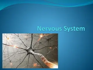
Nervous System and Senses The nervous system consists of two types of cells. Nerve cells = Neurons (Basic unit of nervous system that posses electrical excitability) Glial Cells = Neuroglia (supportive cells, helps to produce myelin sheath)
The parts of a neuron includes the dendrite which receives the impulse (from another nerve cell or from a sensory organ) the cell body (numbers of which side-by-side form gray matter) the axon which carries the impulse away from the cell. Myelin sheath (numbers of which side-by-side form white matter) wrap around the axon and contains Schwann cells . The spaces/junctions between myelin sheaths are called nodes of Ranvier. Diseases which destroy the myelin sheath (demyelinating disorders) can cause paralysis or other problems.
Neuron Structure A neuron has a cell body with mitochondria, lysosomes, a Golgi apparatus, Nissl bodies containing rough endoplasmic reticulum, and neurofibrils.
Nerve fibers include a solitary axon and numerous dendrites. Branching dendrites carry impulses from other neurons (or from receptors) toward the cell body. The axon transmits the impulse away from the axonal hillock of the cell body and may give off side branches. Larger axons are enclosed by sheaths of myelin provided by Schwann cells and are myelinated fibers.
The smallest axons lack a myelin sheath and are unmyelinated fibers. White matter in the CNS is due to myelin sheaths in this area. Unmyelinated nerve tissue in the CNS appears gray. The outer layer of myelin is surrounded by a neurilemma (neurilemmal sheath) made up of the cytoplasm and nuclei of the Schwann cell. Narrow gaps in the myelin sheath between Schwann cells are called nodes of Ranvier.
Types of Neurons & Neuroglial Cells Neurons can be grouped in two ways: 1. structural differences i. Bipolar ii. Unipolar iii. multipolar 2. functional differences i. sensory neurons ii. Interneurons iii. motor neurons 3. Presence of myelin sheath i. Myelinated ii. non-myelinated
Classification of Neurons: According to structural differences ; Bipolar neurons are found in the eyes, nose, and ears, and have a single axon and a single dendrite extending from opposite sides of the cell body. Unipolar neurons are found in ganglia outside the CNS and have an axon and a dendrite arising from a single short fiber extending from the cell body. Multipolar neurons have many nerve fibers arising from their cell bodies and are commonly found in the brain and spinal cord.
According to functional differences ; Sensory neurons (afferent neurons) conducts impulses from the sensory receptors to the CNS (unipolar/bipolar). Interneurons (multipolar) lie within the CNS, transfer and interpret impulses. Motor neurons (multipolar neurons) conduct impulses from the CNS to effectors.
Classification of Neuroglial Cells Neuroglial cells fill spaces, support neurons, provide structural frameworks, produce myelin, and carry on phagocytosis. There are 6 types of glial cells are found in nervous system. 1. Schwann cells are the myelin-producing neuroglia of the peripheral nervous system and other types are components of the central nervous system. 2. Microglial cells are small cells that phagocytize bacterial cells and cellular debris. 3. Oligodendrocytes form myelin in the brain and spinal cord.
5.Astrocytes are near blood vessels and support structures, aid in metabolism, and respond to brain injury by filling in spaces. 6. Ependymal cells cover the inside of ventricles and form choroid plexuses within the ventricles.
Nerve impulse A nerve impulse is an electrical charge that travels down the cell membrane of a neuron s dendrite and/or axon through the action of the Na-K pump. The inside of a neuron s cell membrane is negatively-charged while the outside is positively- charged. When sodium and potassium ions change places, this reverses the inner and outer charges causing the nerve impulse to travel down the membrane.
A nerve impulse is all-or-none: it either goes or not, and there s no halfway. A neuron needs a threshold stimulus, the minimum level of stimulus needed, to trigger the Na-K pump to go and the impulse to travel. A neuron cannot immediately fire again; it needs time for the sodium and potassium to return to their places and everything to return to normal. This time is called the refractory period.
A junction between two nerve cells or a nerve and a muscle cell is called a synapse. In a synapse, various chemicals are used to transfer the impulse across the gap to the next cell. These are collectively known as neurotransmitters, and include such chemicals as dopamine, serotonin, acetylcholine etc.
General Functions of the Nervous System Sensory receptors at the ends of peripheral nerves gather information and convert it into nerve impulses. When sensory impulses are integrated in the brain as perceptions, this is the integrative function of the nervous system. Conscious or subconscious decisions follow, leading to motor functions via effectors.
Nerve Impulse A nerve impulse is conducted as action potential is reached at the trigger zone and spreads by a local current flowing down the fiber, and adjacent areas of the membrane reach action potential.
Impulse Conduction Unmyelinated fibers conduct impulses over their entire membrane surface. Myelinated fibers conduct impulses from node of Ranvier to node of Ranvier, a phenomenon called saltatory conduction. Saltatory conduction is many times faster than conduction on unmyelinated neurons.
All-or-None Response If a nerve fiber responds at all to a stimulus, it responds completely impulse (all-or-none response). by conducting an Greater intensity of stimulation triggers more impulses per second, not stronger impulses.
Nerve Pathways The routes nerve impulses travel are called pathways, the simplest of which is a reflex arc. ReflexArcs A reflex arc includes a sensory receptor, a sensory neuron, an interneuron in the spinal cord, a motor neuron, and an effector. Reflex Behavior Reflexes are automatic, subconscious responses to stimuli that help maintain homeostasis (heart rate, blood pressure, etc.) and responses (vomiting, sneezing, swallowing, etc.). carry out automatic
The knee-jerk reflex (patellar tendon reflex) is an example of a monosynaptic reflex (no interneuron). The interneurons, and motor neurons. withdrawal reflex involves sensory neurons, At the same time, the antagonistic extensor muscles are inhibited.
Brain The brain consists of the cerebrum which is the large, anterior portion. The cerebellum which is the wrinkled-looking, posterior part. The pons which is the closest, larger bulge at the top of the spinal cord. The medulla which is the farther, smaller bulge between the pons and the top of the spinal cord. Then the spinal cord starts after the medulla. Also note under the cerebrum, the optic chiasma, the place where the optic nerves cross to the other side of the brain.
The collectively referred to as the hindbrain. Their functions coordination of maintenance/control of breathing and heart rate. While a stroke in the cerebrum might result in partial paralysis, a stroke in the hind brain is actually, potentially more dangerous because it could knock out coordination of the cerebrum s activities, or worse yet, automatic control of breathing and/or heart beat. cerebellum, medulla, and pons are are involved in homeostasis, movement, and
The midbrain is responsible for receiving and integrating of information and sending/routing that information to other appropriate parts of the brain. The forebrain is composed of the cerebrum and related parts, and functions in pattern and image formation, memory, learning, emotion, and motor control. In addition, the right side functions more in artistic and spatial concepts, while the left side controls speech, language, and calculations. Left-brain stroke would cause paralysis on the right side of the body. It might cause problems with speech while a right-brain stroke is more likely to cause abnormal emotional responses.
Meninges The brain and spinal cord are surrounded by membranes called meninges that lie between the bone and the soft tissues. The outermost meninx is made up of tough, white dense connective tissue, contains many blood vessels, and is called the dura mater. It forms the inner periosteum of the skull bones. In some areas, the dura mater forms partitions between lobes of the brain, and in others, it forms dural sinuses. The sheath around the spinal cord is separated from the vertebrae by an epidural space.
The middle meninx, the arachnoid mater, is thin and lacks blood vessels. It does not follow the convolutions of the brain. Between the arachnoid and pia maters is a subarachnoid space containing cerebrospinal fluid. The innermost pia mater is thin and contains many blood vessels and nerves. It is attached to the surface of the brain and spinal cord and follows their contours.
Spinal Cord The spinal cord begins at the base of the brain and extends as a slender cord to the level of the intervertebral disk between the first and second lumbar vertebrae.
Structure of the Spinal Cord The spinal cord consists of 31segments, each of which gives rise to a pair of spinal nerves. A cervical enlargement gives rise to nerves leading to the upper limbs, and a lumbar enlargement gives rise to those innervating the lower limbs. Two deep longitudinal grooves (anterior median fissure and posterior median sulcus) divide the cord into right and left halves. White matter, made up of bundles of myelinated nerve fibers (nerve tracts), surrounds a butterfly- shaped core of gray matter housing interneurons. Acentral canal contains cerebrospinal fluid.
Functions of the Spinal Cord The spinal cord has two major functions: to transmit impulses to and from the brain, and to house spinal reflexes. Tracts carrying sensory information to the brain are called ascending tracts; descending tracts carry motor information from the brain. The names that identify nerve tracts identify the origin and termination of the fibers in the tract. Many spinal reflexes also pass through the spinal cord.
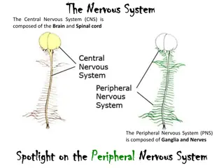
Peripheral Nervous System 1. The peripheral nervous system (PNS) consists of the cranial and spinal nerves that arise from the central nervous system and travel to the remainder of the body. 2. The PNS is made up of the somatic nervous system that oversees voluntary activities, and the autonomic nervous system that controls involuntary activities.
Cranial Nerves 12 pairs of cranial nerves arise from the underside of the brain, most of which are mixed nerves. The 12 pairs are designated by number and name and include the olfactory, optic, oculomotor, trochlear, trigenimal, abducens, glossopharyngeal, vagus, accessory, and hypoglossal nerves. facial, vestibulocochlear,
Spinal Nerves 1. 2. 31 pairs of mixed nerves make up the spinal nerves. Spinal nerves are grouped according to the level from which they arise and are numbered in sequence, beginning with those in the cervical region. Each spinal nerve arises from two roots: a dorsal, or sensory, root, and a ventral, or motor, root. The main branches of some spinal nerves form plexuses. Cervical Plexuses Lie on either side of the neck and supply muscles and skin of the neck. Brachial Plexuses Arise from lower cervical and upper thoracic nerves and lead to the upper limbs. Lumbrosacral Plexuses Arise from the lower spinal cord and lead to the lower abdomen, external genitalia, buttocks, and legs. 3. 4. 5. 6. 7.
Autonomic Nervous System The autonomic nervous system has the task of maintaining homeostasis of visceral activities without conscious effort.
General Characteristics The autonomic nervous system includes two divisions: the sympathetic and parasympathetic divisions, which exert opposing effects on target organs. The parasympathetic division operates under normal conditions. The conditions of stress or emergency. sympathetic division operates under
Autonomic Nerve Fibers In the autonomic motor system, motor pathways include two fibers: a preganglionic fiber that leaves the CNS, and a postganglionic fiber that innervates the effector. Sympathetic Division Fibers in the sympathetic division arise from the thoracic and lumbar regions of the spinal cord, and synapse in paravertebral ganglia close to the vertebral column. Postganglionic axons lead to an effector organ.
Parasympathetic parasympathetic division arise from the brainstem and sacral region of the spinal cord, and synapse in ganglia close to the effector organ. Autonomic Neurotransmitters 1. Preganglionic fibers (PF) of both sympathetic and parasympathetic divisions release acetylcholine. Parasympathetic PF are cholinergic fibers and release acetylcholine. Division Fibers in the 2. Sympathetic postganglionic fibers are adrenergic and release norepinephrine. 3. The effects of these two divisions, based on the effects of releasing different neurotransmitters to the effector, are generally antagonistic.
The sympathetic system. Preganglionic fibers, solid line; post ganglionic fibers, broken lines.
6. Control ofAutonomicActivity a. The autonomic nervous system is largely controlled by reflex centers in the brain and spinal cord. b.The limbic system and cerebral cortex alter the reactions of the autonomic through emotional influence. nervous system
Nervous System Function Somatic NS voluntary muscles and reflexes Autonomic NS visceral/smooth and cardiac muscle Sympathetic NS increases energy expenditure prepares for action These have the opposite effects on the same organs Vs Parasympa thetic NS decreases energy expenditure gains stored energy


 is an")