Understanding the Ossicles of the Ear in Anatomy Studies
Explore the detailed structure of the ear ossicles - the malleus, incus, and stapes - essential for sound transmission. Learn about their components, shapes, and functions in this comprehensive guide from the Department of Anatomy at the Institute of Dental Sciences, Bareilly International University.

Download Presentation

Please find below an Image/Link to download the presentation.
The content on the website is provided AS IS for your information and personal use only. It may not be sold, licensed, or shared on other websites without obtaining consent from the author. If you encounter any issues during the download, it is possible that the publisher has removed the file from their server.
You are allowed to download the files provided on this website for personal or commercial use, subject to the condition that they are used lawfully. All files are the property of their respective owners.
The content on the website is provided AS IS for your information and personal use only. It may not be sold, licensed, or shared on other websites without obtaining consent from the author.
E N D
Presentation Transcript
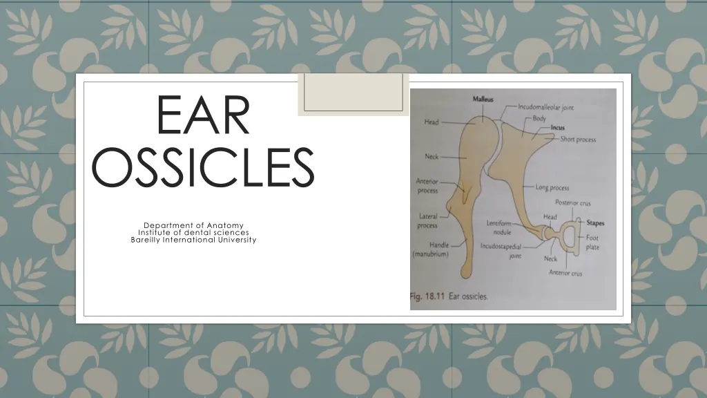
EAR OSSICLES Department of Anatomy Institute of dental sciences Bareilly International University
Malleus It is the largest ossicle which is situated just medial to the tympanic membrane. Malleus is mallet shaped (hammer) and consists of following parts- 1.Head is the larger, rounded upper end of malleus;it lies in epitympanic part and articulates with the incus. 2.Neck It is the constricted part present just below the head. 3.Three proccess -(a). Handle of malleus it is the largest proccess which is directed downwards and is embedded in the medial surface of tympanic membrane. (B) Anterior process- it is a small projection. (C) lateral process it is a conical projection which is attached to the tympanic membrane at the convergence of Anterior & posterior malleolar folds.
Incus It lies between malleus and stapes and present with a body and two processes. (A). Body- it is cubicle in shape.it articulates with head of malleus anteriorly.it forms saddle joint (B). Short process it is a conical projection towards the epitympanic recess posteriorly. (C).long process it extends downwardly from the body and lies parallel to the handle of malleus.the lower end is curved medially and articulates with head of stapes and form Ball & socket joint.
Stapes It is smallest and medial most ossicle. It s shape resembles a stirrup (Rider s feet).and consist of following parts- 1.Head it is small and directed laterally to articulate with incus. 2.Neck-it is seen as a small constricted part under the head,it recieves insertion of stapedius muscles on the posterior surface. 3.Anterior and posterior limbs these arises from the neck and diverge to attach to the base. 4.Base it is also called foot plate of steps,it consists of plate of bone which is reniform (kidney) in shape.the food plate is connected to the fenestra vestibuli (oval window) by an annular ligament.