Understanding Ulcers: Examination and Classification
Explore the detailed examination and classification of ulcers, including descriptions of ulcer floors, bases, and edges. Learn about different types of ulcers, such as traumatic, arterial, venous, neurogenic, infective, tropical, and more. Discover the pathological aspects of non-specific ulcers, specific ulcers, and malignant ulcers.

Download Presentation

Please find below an Image/Link to download the presentation.
The content on the website is provided AS IS for your information and personal use only. It may not be sold, licensed, or shared on other websites without obtaining consent from the author. If you encounter any issues during the download, it is possible that the publisher has removed the file from their server.
You are allowed to download the files provided on this website for personal or commercial use, subject to the condition that they are used lawfully. All files are the property of their respective owners.
The content on the website is provided AS IS for your information and personal use only. It may not be sold, licensed, or shared on other websites without obtaining consent from the author.
E N D
Presentation Transcript
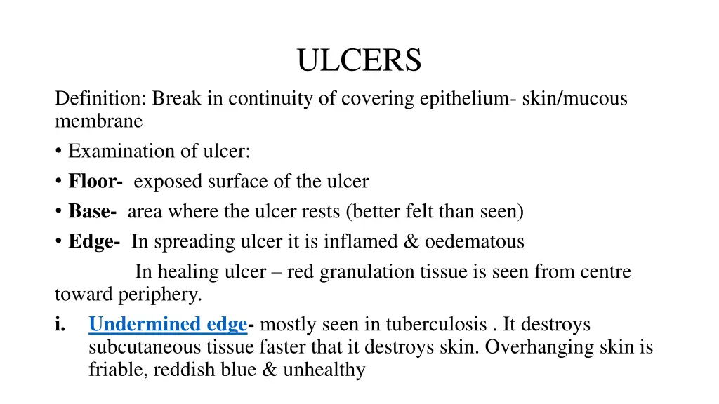
ULCERS Definition: Break in continuity of covering epithelium- skin/mucous membrane Examination of ulcer: Floor- exposed surface of the ulcer Base- area where the ulcer rests (better felt than seen) Edge- In spreading ulcer it is inflamed & oedematous In healing ulcer red granulation tissue is seen from centre toward periphery. i. Undermined edge- mostly seen in tuberculosis . It destroys subcutaneous tissue faster that it destroys skin. Overhanging skin is friable, reddish blue & unhealthy
ii. Punched out edge- seen in gummatous ulcer/ deep trophic ulcer iii. Sloping edge- mostly seen in traumatic/venous ulcer iv. Raised & Pearly white beaded- rodent ulcer. Seen in invasive cellular disease and becomes necrotic at centre. v. Rolled out/Everted edge- characteristic of squamous cell carcinoma / ulcerated adenocarcinoma. The ulcer is a fast growing cellular disease On palpation- induration of edge is characteristic of squamous cell carcinoma/ adenocarcinoma Floor- exposed surface of the ulcer - red granulation tissue (ulcer seems to be healthy and heals) - Pale & smooth granulation tissue (slow healing ulcer) - Wash leathery slough(gummatous ulcer) - Black mass at floor (malignant melanoma)
Base: the area where ulcer rests upon. Induration of base is characteristic of squamous cell carcinoma & Hunterian chancre Classification of ulcer I. Clinically a) Spreading- surrounding area of ulcer is inflamed & floor is covered with slough without any evidence of granulation tissue b) Healing- granulation tissue in floor of ulcer, surrounding skin is not inflamed. Edge shows bluish outline of growing epithelium. Slight serous discharge is seen. c) Callous- pale granulation tissue in floor of ulcer. Induration of base, edge & surrounding skin. Shows no tendency to healing.
II: Pathologically A. Non specific ulcers B. Specific ulcers C. Malignant ulcers Non specific ulcers a) Traumatic ulcer- i. Mechanical- dental ulcer on tongue from jagged tooth ii. Chemical- application of caustics iii. Physical- electrical. X-ray burns b) Arterial ulcer- atherosclerosis , buerger s disease & Raynaud s disease c) Venous ulcer- post phlebitic limb
d) Neurogenic ulcer e) Infective ulcer- Pyogenic & Bairnsdale ulcers f) Tropical ulcer- infection is by Vincent s organisms(bacteroides fusiformis). Small abrasion may cause ulcer. Ulcers associated with malnutrition, anaemia, avitaminosis are included. g) Cryopathic ulcer- ulcers due to chilblains & cold injury h) Martorell s ulcer- hypertensive ulcer i) Bazin s ulcer- erythrocyanoid ulcer j) Diabetic ulcer k) Miscellaneous- polycythemia, leukaemia, systemic sclerosis, ulcerative colitis, poliomyelitis, arteriovenous fistula, collagen disorder, chronic lymphoedema, acholuric jaundice
B) Specific ulcers- tuberculosis, syphilis, soft sore, actinomyces, meleneys ulcer C) Malignant ulcers- Epithelioma, rodent ulcer, malignant melanoma Traumatic Ulcer- situated anywhere in the body. More commonly in bony prominences- shin, malleoli, back of heel. Small, painful & circular ulcers Eg- Footballer s ulcer- repetitive trauma(usually due to staphylococcal infection) They heal quickly unless intervened by ischemia/infection.
Arterial ulcer: - Caused due to inadequate skin circulation - Parts subjected to repeated pressure & trauma - Mostly due to peripheral arterial disease & poor peripheral circulation - Atherosclerosis is commonest condition(seen in old people) - Usually on anterior & lateral aspect of legs, toes, dorsum of foot Buerger s disease- Age : 20-40yrs. Patches of dry gangrene are present along arterial ulcer. - Pain is the main complaint - These ulcers tend to be punched out & destroy whole skin & deep fascia(unlike venous ulcer)
Venous ulcer: Abnormal venous hypertension. - Usually seen in lower 1/3rdof leg, ankle, dorsum of foot. - Various terms used are post thrombotic ulcer, gravitational ulcer, varicose ulcer Neurogenic ulcer: Repeated injury/pressure in an area which has lost appreciation of pain Eg- diabetes, alcoholic, peripheral neuritis, tabes dorsalis, spina bifida, leprosy, paraplegia, syringomyelia. - Commonly seen on heel & ball of foot when patient is ambulatory & on buttocks & heel when patient is non ambulatory. - This ulcer starts as a callosity under which suppuration takes place> pus comes out> central hole form in the ulcer> burrows through muscle & bone - Trophic ulcer: impairment of nutrition , inadequate blood supply & neurogenic deficit
Tropical ulcer: - Edge is slightly raised & exudes copious serosanguineous discharge - Retains same size for month s/years - In some cases- destroys surrounding skin tissue & spreads widely - Infection by Vincent s organism-abrasion/ break in continuity of skin > papule with surrounding inflammation & oedema. - Pain is important symptom - Gradually pustules develop & burst in 2-3 days - Copious serosanguineous discharge - Ulcer becomes indolent & refuses to heal for month s/ years.
Cryopathic ulcers: i) Chilblains(perniosis)- lower extremities exposed to intense cold- blisters , ulceration. - Lesion starts as red tender spot then pruritic swelling - Blisters also form - Blisters burst to form ulcers - Ulcers are superficial ii) Cold injury(frost bite): - Any part of the body exposed to wet cold below freezing point, ischaemic changes occur in skin & subcutaneous tissue. - Ischaemia> arteriolar spasm& stasis of blood in capillaries> Freezing of tissues & denaturation of intracellular protein. - Later on gangrene ensues(full thickness of skin)
Martorells ulcer: - Described by Martorell 1945 - Patients above 50 yrs of age who are hypertensive & atherosclerotic - Local patch on back/outer side of calf suddenly necroses & sloughs out leaving a punched out ulcer to deep fascia. - Pain is severe & prevents sleeping. Condition may be bilateral Pathology- sudden obliteration of end arterioles of skin which is already having sparse arterial supply with atherosclerosis
Bazins ulcer: - Associated with erythrocyanosis frigida - Thick ankles with abnormal amount of subcutaneous fat with abnormally poor arterial supply to ankle skin - Post tibial arteries/peroneal arteries- abnormally small/ having ischaemic changes - Sensitive to temperature changes - Cold season- skin is blue, cold/tender - Hot season- Hot oedematous, swollen, painful (reactive hyperaemia) - Ulcers are small & multiple Surgical intervention- sympathectomy
Diabetic Ulcer: - Slight injury to glucose laden tissue > chronic infection & ulcer formation - May be precipitated by ischaemia/diabetic atherosclerosis/peripheral neurosis Miscellaneous: Cortisone ulcers, acholuric jaundice Tuberculous ulcer: - Such ulcers develops due to bursting of cold abscess - Matted tuberculous lymph node - Tuberculosis of bone /joint - Submucosal lesion- intestinal. Tongue tuberculosis.
Characteristics: Size & shape- oval in shape with irregular crescentic border Number- multiple Edge- reddish blue & undermined Depth- shallow Pain- slight+ Floor- pale granulation tissue is seen at floor with variable amount of discharge Base- slight induration Lupus vulgaris: - Cutaneous tuberculosis occurs most commonly in face & hand. Victim is usually child & young adults - Heals at centre & remains active at periphery & thus gradually spreads like wolf
Syphilitic ulcers: I. Primary syphilis- hard chancre/Hunterian chanre - Develops at site of entry of treponema; 3-4 weeks of exposure - Sites- external genitalia Other sites- lip, tongue, nipple, perianal region Characterisitcs: - single, painless, indurated base(button) - Regional lymph nodes; firm discrete, painless
II. Secondary Mucous patches- white patches of sodden thickened epithelium Snail track ulcers- small, round & superficial erosions coalesce to form narrow , curved & shallow ulcers Condylomata lata- fleshy wart like growths seen at mucocutaneous junction- mouth, anus & vulva - Round, white, flat with hypertrophied epithelium - Enlarged lymph nodes- painless (epitrochlear & suboccipital)
III. Teritiary Gummatous ulcer- granulation tissue with central necrosis Sloughing of the necrotic tissue- punched out indolent edge & painlessness Floor- yellowish grey gummatous tissue Healing- silvery tissue paper scar WR, Kahn & VDRL : +ve. Lymph nodes are not enlarged unless secondarily infected Subcutaneous tissue- tibia, sternum, ulna, skull Scrotum- anterior surface in relation to testis Tongue(occasionally)
Soft chancre/chancroid(ducreys): - Caused by gram ve Haemophilus Ducreyi - Contagious disease - In 3-5 days of exposure ; multiple acute sores develop on external genitalia - Painful, gradually become pustular & ulcerate to form soft sores - Regional lymph nodes are enlarged & tender (acute lymphadenitis) - If suppuration occurs nodes become matted to form a fluctuant swelling with abscess(bubo) - Overlying skin becomes red
Actinomycosis: - Multiple ulcers - Indurated area> nodules appear> soften & ulcerate in various places - Bluish skin surrounding ulcers Diagnosis: collecting discharge pus in sterile tube; presence of granules sulphur granules , gram +ve mycelia( peripheral filaments radiates from central part of granules. i. Facio- cervical (commonest) ii. Thorax iii. Right iliac fossa iv. Liver
Meleneys ulcer: - Post operative wounds after operation for perforated viscus/drainage of empyema thoracis. - Rare- dorsum of hand - Symbiotic action- non haemolytic streptococci & haemolytic staphylococcus aureus - Undermined ulcer with lot of granulation tissue in floor. Surrounded by deep purple zone, in turn surrounded by outer zone of erythema - Painful, toxaemic & general condition deteriorates without treatment.
Therapeutics Graphites: scabby eruptions on scalp , face, bends of joints. Corners of mouth, and eyes are cracked, bleeding & oozing a gluey, tenacious discharge. Fissured eczema. Lycopodium- dry scaly eruptions Calcarea carbonica: eczema on scalp which extends to face. Crusts are white on awakening. In morning the child will scratch furiously Arsenicum: thickened skin as in chronic eczema. Itching , burning & swelling. Papules, nettle rash & swelling. Burning sensitive ulcers with offensive discharge. Bovista: Baker s & grocer s itch; eruptions on back of hands Sepia: Dry desquamation. Ringworm.
Clematis: rawness, worse washing, moist eruption Sulphur: aggravation from washing. Scratching makes the part burn intensely. Skin is rough, coarse and measly, soreness in fold of skin & a tendency to pustular eruption. Dryness with heat of scalp, with intense itching, especially at night & scratching causes soreness; wetting makes it burn. Eruptions of yellow crusts Selenium: itching in folds of skin and about ankle joints. Antimonium crudum: thick callosities of skin. Honey coloured crusts on heads of children; cracking in nostrils & corners of mouth Thuja: warts and eczema following vaccination Natrum muriaticum: dry scaly or herpetic eruptions of little blisters in bends of joints. Fever blisters. Moist eczema without much itching. Eczema with thick scabs oozing pus. Urticaria with itching about the joints when occurring with intermittent fever worse at sea shore.
Kreosote: eruption on extensor surface of joints Berberis aquifolia: scaly pustular eruption on face Hydrocotyle: great dryness and desquamation of the epidermis. Acne rosacea. Dry and impoverished skin, cold hands and feet Petroleum: eczema with thick scabs, oozing pus and rhagades; skin is harsh and dry, finger tips crack and hands chap. Especially suitable to eczema behind ears. Mezereum: Itching worse when the patient is warmed or wrapped up. Small vesicles with terrible itching , secretion dries quickly, producing scabs from beneath which thick pus oozes. Herpes zoster Rhustoxicodendron: right sided herpes zoster with extensive vesication accompanied by rheumatic pains. Itching and tingling sensation. Skin is oedematous and swollen. All symptoms are worse at night, damp weather and winter
Apis: burning and stinging pain with oedema. Cantharis: large blisters with smarting and burning Anacardium: small blisters with umbilicated center. Eruptions itching excessively and burns Dolichos: itching without eruptions. Useful in senile pruritis. Worse at night. Worse across shoulders. Psorinum: herpetic eruptions with itching, worse when getting warm in bed. Skin is dirty greasy, unwashed in appearance. Tinea capitis. Increased secretions or boils remaining after itch. Oleander: skin eruptions with gastro enteric troubles; skin is very sensitive. Slight friction causes chafing and soreness, especially about the neck, scrotum and thighs. Crusta lactea on scalp and back. Great itching; scratching relieves at first. But parts become very sore.
Vinca minor: eczema of scalp and face. Matted hair and offensive odor. Crust is formed and discharge is retained underneath; causing hair to fall out or mat together- plica polonica Viola tricolor: crusta lactea and impetigo in children. Crusts with copious exudation and is accompanied with offensive urine. Staphysagria: eruptions worse on occiput, eczematous eruption on ears, yellow scabs. Scratching changes place of itching. Eruptions form figwarts or condylomata. Sickly children after abuse of mercury. Ranunculus bulbosus: vesicular eruptions along course of nerves. Vesicles filled with serum & burn greatly. Large blisters form on raw surface. Herpes zoster. Pemphigus in new born. Thickening of skin into yellow hard horny scabs.
Nitric acid: ragged zig zag , often raised edges i. Profuse granulations, proud flesh ii. Vesicular bleeding easily where touched iii. Splinter like pains in them . Sycotic excrescences. Sepia: brownish spots on skin. Herpetic eruptions about knees and ankles. Bends of joints and behind the ears; at first dry, it becomes moist and discharges copiously. Ringworm , herpes circinatus Hepar sulph: moist eruptions in folds of skin and itching in bends of joints. Skin is extremely sensitive and suppurates easily, and pimples form around ulcers. Humid eczema of scalp, sore and sensitive to touch. Eczema of scrotum and genital organs, boils. After abuse of ointments containing zinc or mercury.
Kali bichromicum- eczema capitis and moist eczema, when chronic and obstinate in character. Useful remedy in acne & sycotic manifestations.


 Neurogenic ulcer")
 Specific ulcers- tuberculosis, syphilis, soft sore,")


:")