Understanding X-ray Generation Process: Bremsstrahlung and Characteristic Radiation
Learn about the production of X-rays through Bremsstrahlung and Characteristic radiation. Discover how high-speed electrons interact with target nuclei in X-ray tubes, resulting in the emission of photons with varying energy levels.

Download Presentation

Please find below an Image/Link to download the presentation.
The content on the website is provided AS IS for your information and personal use only. It may not be sold, licensed, or shared on other websites without obtaining consent from the author. If you encounter any issues during the download, it is possible that the publisher has removed the file from their server.
You are allowed to download the files provided on this website for personal or commercial use, subject to the condition that they are used lawfully. All files are the property of their respective owners.
The content on the website is provided AS IS for your information and personal use only. It may not be sold, licensed, or shared on other websites without obtaining consent from the author.
E N D
Presentation Transcript
Production of x rays Bremsstrahlung radiation Characteristic radiation
Bremsstrahlung radiation These interactions are the primary source of x ray photons from x ray tube They are produced by sudden stopping or slowing of high speed electrons at target When electrons from filament strike target, x ray photons are created if electrons hit a target nucleus directly or if their path takes them close to a nucleus If electron directly hits nucleus, all its kinetic energy is tranformed into single x ray photon Energy of resultant photon is equal to energy of kV applied across x ray tube at that instant of its passage
Most electrons have near or wide misses with atomic nuclei Here vely charged high speed electron is attracted toward +vely charged nuclei and losses some of its velocity This deceleration causes electron to lose some kinetic energy, which is given off in the form of many new photons The closer the high speed electron approaches the nuclei, greater is the electrostatic attraction on electron, the braking effect, and the energy of the resulting bremsstrahlung photons
These interactions generate x ray photons with a continuous spectrum of energy: reasons being: 1. Continuously varying voltage difference b/w target and filament, characteristics of half wave recitification, causes the electron striking the target to have varying levels of KE 2. The bombarding electrons pass at varying distances around tungsten nuclei and are thus deflected to varying extents. As a result, they give up varying amounts of energy in the form of radiation 3. Most electrons participate in this interactions. Consequently, electron carries differing amounts of energy at the time of each interaction with tungsten nucleus results in generation of x ray photons
Characteristic radiation It occurs when an electron from filament displaces an electron from a shell of tungsten target atom, thereby ionising the atom Higher energy electron in an outer shell of tungsten atom is quickly attracted to the void in deficient inner shell When outer shell electron replaces displaced electron, a photon is emmitted with an energy equivalent to the difference in the two orbital binding energies
Characteristic radiation from K shell occurs only above 70 kVp with a tungsten target and occurs as discrete increments compared to Bremsstrahlung radiation Energies of characteristic photons are a function of the energy levels of various electron orbital levels and hence are characteristic of target atoms Characteristic radiation is only a minor source of radiation from an x ray tube
Factors controlling the X ray beam Exposure time: when exposure time is doubled, no of photons generated at all energies in x ray emmission spectrum is doubled Changing the time controls the quantity of the exposure, the no of photons generated
Tube current As mA setting is increased, more power is applied to filament, which heats up and releases more electrons that collide with the target to produce radiation Quantity of radiation produced by x ray tube is directly proportional to tube current and the time the tube is operated It is expressed as product of time and tube current Quantity of radiation remains constant regardless of variations in mA and time as long as their product remains constant
Tube voltage Increasing the kVp increases the potential difference b/w cathode and anode, thus increasing the energy of each electron when it strikes the target This results in increased efficiency of conversion of electron energy into x ray photons and thus an increase in: 1. The no of photons generated 2. Their mean energy 3. Their maximal energy
Half value layer It s a useful way to characterize the penetrating quality of an x ray beam It is the thickness of an absorber, such as aluminium, required to reduce by one half the no of x ray photons passing through it As the average energy of an x ray beam increases, so does its HVL
Filtration The x ray photons carry different energies and photons with sufficient energy can penetrate through atomic structures and reach image receptor Low energy photons contribute to pt exposure and thus should be removed This is accomplished by placing an aluminium filter in the path of beam Al preferentially removes many low energy photons with less effect on high energy photons that are able to penetrate the film
Amount of filteration required for particular x ray machine, kVp and inherent filteration of tube and its housing must be considered Inherent filteration consists of materials that x ray photons encounter as they travel from focal spot on target to form a usable beam outside tube enclosure These include, glass wall of x ray tube, insulating oil, and barrier that prevents oil from escaping through the x ray port Inherent filteration ranges from 0.5 to 2mm of aluminium
Total filteration is sum of inherent filteration plus any added external filteration External filteration supplied in the form of aluminium disks placed over the port in the head of x ray machine Governmental regulations require total filteration to equivalent of 1.5mm aluminium to 70 kVp and 2.5 mm for all higher voltages
Collimation A collimator is a metallic barrier with an aperature in the middle used to reduce the sixe of x ray beam and therefore volume of irradiated tissue within the pt Round and rectanglar are frequently used Dental x ray beams are usually collimated to a circle 23/4 inches/7cm in diameter Round collimator is a thick plate of r opaque material, usually lead with a circular opening centered over the port in x ray tube head through which x ray beam emerges
Rectangular collimators further limit the size of the beam to just larger than the x ray film Use of collimation also improves image quality Tissues in the path absorbs about 90% of photons and 10% of photons pass through the pt and reach the films Many absorbed photons generate scattered radiation within the exposed tissues by comptom scattering These scattered photons travel in all directions and some reach the film and degrade image quality Collimating the beam to reduce exposure area and thus no of scattered photons reaching the film can minimize the detrimental effect of scattered radiation on images
Inverse square law Intensity of an x ray beam at a given point depends on the distance of the measuring device from focal spot For given beam, intensity is directly proportional to square of the distance from the source Reason for this decrease in intensity is that the x ray beam spreads out as it moves from the source Relationship: I1/I2 = (D2)/(D1)
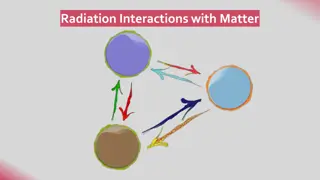
Interactions of x rays with matter intensity of an x ray beam is reduced by interaction with the matter it encounters Attenuation results from interactions of individual photons in the beam with atoms In the absorber X ray photons are either absorbed or scattered out of the beam
Coherent scattering Syn: classical, elastic or Thompson scattering It occurs when a low energy incident photon passes near an outer electron of an atom Incident photon interacts with the electron by causing it to become momentarily excited at the same frequency as the incoming photon Incident photon ceases to exist Excited electron then returns to ground state and generates another x ray photon with the same frequency and energy as in the incident beam
The secondary photon is emitted at an angle to the path of the incident photon This interaction accounts for 8% of total no of interactions per exposure It contributes very little to film fog coz the total quantity of scattered photons is small and its energy level is too low for it to reach the film
Photoelectric absorption This occurs when an incident photon collides with a bound electron in an atom of the absorbing medium At this point, incident photon ceases to exist Electron is ejected from its shell and becomes a recoil electron/photoelectron Kinetic energy imparted to recoil electron is equal to the energy of incident photon minus that used to overcome the binding energy of the electron Absorbing atom is now ionised coz it has lost an electron
This electron deficiency is instantly filled with a release of characteristic radiation Recoil electrons ejected travel only short distances in the absorber before they give up their energy through secondary ionisations Most interactions occur in K shell coz the density of electron cloud is greater in this region and higher probability of interaction exists About 30% of interactions are by photoelectric process
Compton scattering It occurs when a photon interacts with an outer orbital electron Incident photon collides with an outer electron, which receives kinetic energy and recoils from the point of impact The path of the incident photon is deflected by its interaction and is scattered from the site of the collision Energy of the scattered photon equals the energy of the incident photon minus the sum of kinetic energy gained by recoil electron and its binding energy
This results in the loss of an electron and ionisation of the absorbing atom Scattered photons continue on their new paths, causing further ionizations Recoil electrons also give up their energy by ionizing other atoms About 62% of photons are absorbed by this process Scattered photons travel in all directions Higher the energy of incident photon, greater the probability that the angle of scatter of secondary photon will be small and its direction forward
30% of scattered photons formed during exposure will exit through pts head It is disadvantageous coz it causes film darkening