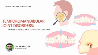
Learn about the unique structure and function of the temporomandibular joint, which involves multiple bones such as the mandible, maxillae, temporal bone, and more. Discover the bony composition, articular surfaces, and components like mandibular condyles and ligaments that make up the TMJ.
Please find below an Image/Link to download the presentation.
The content on the website is provided AS IS for your information and personal use only. It may not be sold, licensed, or shared on other websites without obtaining consent from the author. If you encounter any issues during the download, it is possible that the publisher has removed the file from their server.
You are allowed to download the files provided on this website for personal or commercial use, subject to the condition that they are used lawfully. All files are the property of their respective owners.
The content on the website is provided AS IS for your information and personal use only. It may not be sold, licensed, or shared on other websites without obtaining consent from the author.
E N D
Presentation Transcript
Dr Sarath Babu V Professor cum Principal Dept. of Sports Physiotherapy MGM Institute Of Physiotherapy Chh. Sambhajinagar
The temporomandibular (TM) joint is unique in both structureand function. Multiple bones merge to form the structure and contribute to the function of the TM joints. These bones include the mandible, maxillae, temporal, zygomatic, sphenoid, and hyoid bones. The proximal or stationary segment of the TM joint is the temporal bone.
A cross-sectional lateral view of the TM joint shows the fibrocartilage-covered bearing surfaces condyle of the mandible and the articulareminence. The TM disc divides the articulation into an upper joint and a lower joint, each with its own synovial lining. The anterior and posterior attachments capsule to the disc are shown. load- the on of the joint
This is a synovial joint of condylar variety. Articular surfaces- A. upper part - a) Articular eminence, b) Ant. part of mandibular fossa. B. inferior surface - a) head of the mandible.
The fibers near the surface of the articular covering are aligned in a parallel arrangement to facilitate gliding of the joint surfaces. The presence of fibrocartilage rather than hyaline cartilage is significant because fibrocartilage can repair and remodel itself.
Typically fibrocartilage is present in areas that have to withstand repeated and high-level stress. The TM joints are subjected to the repetitive stress of jaw motions as well as to tremendous bite forces, which have been measured at 597 N for women and 847 N for men.
The mandible is the largest of the facial bones and is highly mobile. The mandible is arch-shaped and consists of a condyle at each posterior superior portion. Each mandibular condyle has a medial and lateral pole, and the lateral pole is readily palpable
Accessory Joint Structures The incongruence of the TM joint is addressed by a unique articulardisc. The inferior TM joint is formed by the mandibular condyle and the inferior surface of the disc and functionsas a simple hinge joint The superior TM joint is larger than the inferior joint and is formed by the articular eminence of the temporal bone and the superior surface of the disc; it functionsas a gliding joint.
Articular Disc A cross-section of the temporomandibular joint showing articulardisc. The thickness of the disc varies from anteriorly to 1 mm in the center to posteriorly. the 2 mm 3 mm
Capsule and Ligaments The elasticity of the joint capsule and ligaments determines the available motion at the TM joint in all planes
capsule The capsule is a fibrous membrane that surrounds the joint and incorporates the articular eminence. It attaches to the articular eminence, the articular disc and the neck of the mandibular condyle. The portion of the capsule superior to the disc is quite lax, whereas the portion of the capsule inferior to the disc is taut. Lack of capsular strength anteriorly predisposes anterior dislocation of mandibular condyle
The anterior portion of the disc attached to joint capsule & lateral pterygoid.This ant. Attachment restricts posterior translation. The posterior portion attached to retrodiscal tissue superior & inferior lamina. Sup. Lamina attached posteriorly at tympanic plate allows ant. Translation during mouth opening & repositioning due to its elastic property during mouth closing. Inf. Lamina limits forward translation
Ligaments The primary ligaments of the TM joint are the TM ligament, the stylomandibularligament, and the sphenomandibularligament.
TM ligament The TM ligament is a strong ligament composed of twoparts, an outer oblique element and an inner horizontal element. . The primary function of the TM ligament is to stabilize the lateral portion of the capsule. Neither band of the TM ligament limits forward translation of the condyle or disc, but they do limit lateral Displacement.
The outer oblique element attaches to the neck of the condyle and eminence It serves as a suspensory ligament downward and motion of the mandible, as well as limiting rotation of the condyle mandibulardepression The component of the ligament is attached to the lateral pole of the condyle and posterior portion of the disc and to the articulareminence. Its fibers arealigned horizontally posterior motion condyle. Limiting the translation of the condyle protects the retrodiscal pad inner horizontal the articular and limits posterior to resist the of during posterior
stylomandibular ligament The stylomandibular ligament is the weakest of the three ligaments and is considered a thickened part of the parotid sheath joining the styloid process to the angle of the mandible. function of this ligament as limiting the protrusion of the mandible.
sphenomandibular ligament The sphenomandibular ligament is described as the strong ligament that is the swinging hinge from which the mandible is suspended. it serves to protect the mandible from excessive anterior translation.
The sphenomandibular ligament attaches to the spine of the sphenoid bone and to the middle surface of the ramus of the mandible. sphenomandibular ligament also has continuity with the disc medially. the sphenomandibular ligament serves as an accessory ligament and, in concert with the TM ligament, provides structural support for the TM joint.
Joint Kinematics The TM joint is one of the most frequently used and mobile joints in the body. It is engaged during mastication, swallowing, and speaking. Most of the time, the TM joint movements occur without resistance from chewing or contact between the upperand lowerteeth. However, as a third-class lever, the TM joint is designed to maintain its structure in spite of significant forces acting on it.
Osteokinematic motions include mandibular depression, elevation, protrusion, retrusion, left and right lateral excursions Arthrokinematic movements involve rolling, anterior glide, distraction, lateral glide.
Depression- lateral pterygoid mainly Elevation- masster,temporalis,medial petygoid of both sides. Protrusion- lateral and medial pterygoid. Retraction- posterior fibres of temporalis. Lateral or side to side movement eg.turning chin to left side- left lateral pterygoid and right medial pterygoid.
To accomplish mandibular depression and elevation, the mandibularcondyle must roll and glide. During rotation, the mandibular condyle spins relative to the inferiorsurfaceof thedisc in the lower joint . Translation occurs in the upper joint between the disc and the articular eminence.
Depression(mouth opening) Normal mouth opening is done by Rotation in lower joint between disk & condyle with pure ant. Rotation of condyle on disk gliding or translation anteriorly & inferiorly in upper joint between disk & articular eminence. Normal mandibular depression range of motion is 40 to 50 mm Mastication requires approximately 18 mm of mandibular depression.
. Elevation (mouth closing)- reverse of depression post. Translation & post. Rotation Mandibular elevation is the reverse of mandibular depression. The mandibular condyle rotates posteriorly on the disc in the lower joint, and the condyle-disc complex translates posteriorly in the upper joint.
Mandibular Protrusion and Retrusion Maximum protrusion, teeth should be in front of the upperteeth. mandibular the lower
Mandibular protrusion and retrusion occur in the upper TM joint. The condyle-disc complex translates in an anterior inferior direction, following the downward slope of the articular eminence, during protrusion and returns along a posterior superior path. Protrusion is an important component necessary for maximal mandibular depression. Retrusion is an important component of mandibular elevation from a maximally depressed mandible..
Mandibular Lateral Excursion With deviation mandible to the right, the midline of the lower teeth should move the full width of the right uppercentral incisor. normal lateral the of
Muscles The muscles acting on the TM joint are divided into primary and secondary muscle groups
Primary Muscles Temporalis Masseter Lateral pterygoid, and medial pterygoid.
Secondary Muscles The secondary muscles are smaller than the primary muscles and consist of the suprahyoid and infrahyoid groups. The digastric, geniohyoid, mylohyoid, and stylohyoid comprise the suprahyoid group
Coordinated Muscle Actions Mandibular depression occurs from the concentric action of the bilateral digastric muscles in conjunction with the inferior portion of the lateral pterygoid muscles. Mandibular elevation results from the collective concentric action of the bilateral masseter, temporalis, and medial pterygoid muscles.
The most common disorder of the TMJ is disc displacement. The most common cause of TMJ pain is myofascial pain dysfunction syndrome, primarily involving the muscles of mastication. Internal derangements is defined as an abnormal relationship of the disc to any of the other components of the TMJ. Disc displacement is an example of internal derangement
Degenerative joint disease, otherwise known as osteoarthritis is the organic degeneration of the articular surfaces within the TMJ. TMJ pain remains one of the most reliable diagnostic criteria for temporal arthritis.


 joint is unique in")


 –")
- reverse of depression post.")