Unit V DNA sequencing
DNA sequencing is the essential process of deciphering the order of nucleotides in DNA, which involves methods to determine the sequence of adenine, guanine, cytosine, and thymine bases. This crucial technique is employed in various fields of research and diagnostics to unravel genetic information and understand biological processes at a molecular level.

Download Presentation

Please find below an Image/Link to download the presentation.
The content on the website is provided AS IS for your information and personal use only. It may not be sold, licensed, or shared on other websites without obtaining consent from the author.If you encounter any issues during the download, it is possible that the publisher has removed the file from their server.
You are allowed to download the files provided on this website for personal or commercial use, subject to the condition that they are used lawfully. All files are the property of their respective owners.
The content on the website is provided AS IS for your information and personal use only. It may not be sold, licensed, or shared on other websites without obtaining consent from the author.
E N D
Presentation Transcript
Unit V DNA sequencing DNA sequencing is the process of determining the nucleic acid sequence the order of nucleotides in DNA. It includes any method or technology that is used to determine the order of the four bases: adenine, guanine, cytosine, and thymine. The rapid speed of sequencing attained with modern DNA sequencing technology has been instrumental in the sequencing of complete DNA sequences, or genomes, of numerous types and species of life, including the human genome and other complete DNA sequences of many animal, plant, and microbial species.
Frederick Sanger, a pioneer of sequencing. Sanger is one of the few scientists who was awarded two Nobel prizes, one for the sequencing of proteins, and the other for the sequencing of DNA.
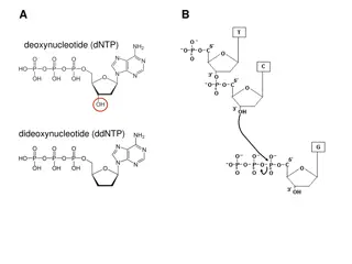
Enzymatic Chain Termination Method (Sanger sequencing or dideoxy method) Developed by Sanger and coworkers in 1977. This method is based on the DNA polymerase dependent synthesis of a complementary DNA strand in the presence of dNTPs and dideoxynucleotides (ddNTPs) that serve as terminators. The DNA synthesis reaction is randomly terminated whenever a ddNTP is added in the Growing oligonucleotide chain, resulting a truncated products of varying lengths with an appropriate ddNTP at their 3 -terminus. The products are separated by using capillary gel electrophoresis and its terminal ddNTPs are used to reveal the DNA sequence of the strand.
Nowadays, Automated sequencing instruments combine sequencing with fluorescently labeled primers or dideoxy chain terminators with polyacrylamide gel electrophoresis and computer data capture. Sanger sequencing is usually automated.
Sanger DNA sequencing is widely used for research purposes like: 1. Targeting smaller genomic regions in a larger number of samples 2. Sequencing of variable regions 3. Validating results from next-generation sequencing (NGS) studies 4. Verifying plasmid sequences, inserts, mutations 5. HLA typing 6. Genotyping of microsatellite markers 7. Identifying single disease-causing genetic variants
MaxamGilbert sequencing Maxam Gilbert sequencing is a method of DNA sequencing developed by Allan Maxam and Walter Gilbert in 1976 1977. This method is based on nucleobase-specific partial chemical modification of DNA and subsequent cleavage of the DNA backbone at sites adjacent to the modified nucleotides. Maxam-Gilbert sequencing requires radioactive labeling at one 5' end of the DNA and purification of the DNA fragment to be sequenced. Chemical treatment then generates breaks at a small proportion of one or two of the four nucleotide bases in each of four reactions (G, A+G, C, C+T). The concentration of the modifying chemicals is controlled to introduce on average one modification per DNA molecule. Thus a series of labeled fragments is generated, from the radiolabeled end to the first "cut" site in each molecule. The fragments in the four reactions are electrophoresed side by side in denaturing acrylamide gels for size separation. To visualize the fragments, the gel is exposed to X-ray film for autoradiography, yielding a series of dark bands each corresponding to a radiolabeled DNA fragment, from which the sequence may be inferred.
4 different chemicals cleave DNA at four different positions; hydrazine and hydrazine NaCl selectively attack pyrimidine nucleotides while dimethyl sulfate and piperidine attack purine nucleotides. Take a look at the chemistry here. 1. Hydrazine: T + C 2. Hydrazine NaCl: C 3. Dimethyl sulfate: A + G 4. Piperidine: G Denatured DNA of equal volume is added to 4 separate tubes containing these 4 different chemicals. Afterward, sample incubation is performed followed by gel electrophoresis using PAGE- polyacrylamide gel electrophoresis. The results of chemical cleavage are shown in the illustration given below. To enable visualization autoradiography analysis is carried out. The presence of 32P on the DNA allows us to determine fragments generated by the cleavage.
Pyrosequencing is a method of DNA sequencing (determining the order of nucleotides in DNA) based on the "sequencing by synthesis" principle, in which the sequencing is performed by detecting the nucleotide incorporated by a DNA polymerase. Pyrosequencing relies on light detection based on a chain reaction when pyrophosphate is released. Hence, the name pyrosequencing.
The first full DNA genome to be sequenced was that of bacteriophage X174 in 1977. In 1995, Venter, Hamilton Smith, and colleagues at The Institute for Genomic Research (TIGR) published the first complete genome of a free-living organism, the bacterium Haemophilus influenzae.