University of Basrah College of Nursing Health Promotion Course
Dive into the crucial components of health assessment focusing on examination and inspection techniques in nursing practice. Explore the systematic process of assessing general and specific health indicators, including vital signs and abdominal examination.

Download Presentation

Please find below an Image/Link to download the presentation.
The content on the website is provided AS IS for your information and personal use only. It may not be sold, licensed, or shared on other websites without obtaining consent from the author.If you encounter any issues during the download, it is possible that the publisher has removed the file from their server.
You are allowed to download the files provided on this website for personal or commercial use, subject to the condition that they are used lawfully. All files are the property of their respective owners.
The content on the website is provided AS IS for your information and personal use only. It may not be sold, licensed, or shared on other websites without obtaining consent from the author.
E N D
Presentation Transcript
University of Basrah College of Nursing Health Promotion Course Fourth Year Students First Semester 2023
Lecture no. 7 Components of the Health Assessment Lecturer Dr. FirasA. Jasim (MBChB-ABMS-FM)
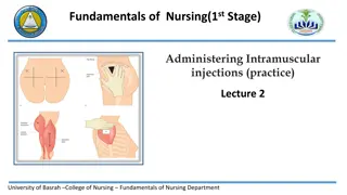
Components of the Health Assessment 2/ Examination A/ General examination - Examination of the exposed parts of the body (head, neck, hands and feet) and examination of the vitals (Bp, pulse rate, respiratory rate and the body temperature).
-General inspection: - Age - Built - Position in bed - Expression - Jaundice - Pallor - Cyanosis - Clubbing of fingers - Leg Oedema - Cervical lymphadenopathy - Examination of the head and neck - Examination of the hands and feet - Vital signs : Pulse, Temperature, Respiratory rate and Blood pressure.
B/ Systematic examination Inspection. Palpation. Percussion. Auscultation.
Abdominal examination The abdomen is bordered superiorly by the costal margins, inferiorly by the symphysis pubis and inguinal ligaments and laterally by the flunks. The abdomen divided into 9 regions or quadrants commonly used to describe abdominal findings. Upper regions [right hypochondriac, left hypochondriac and epigastric]. Middle region[ right lumber, left lumber and umbilical]. Lower region [right iliac fossa, left iliac fossa and suprapubic region].
In order to do abdominal examination we have to take the permission from the patient and we put the patient in supine position and we expose the patient gently from the nipple to the mid thigh, because : some of abdominal organs lies below the rib cage e.g., liver, spleen. and the external genitalia are part of the abdomen.
Inspection of abdomen 1/ From the foot of the bed: We mention the symmetry and shape of the abdomen: # Asymmetry may be caused by organomegally like hepatomegallyor splenomegally or because of the presence of an abdominal mass like large ovarian cyst or large hernia.
# The shapeof the abdomen may be one of the followings:- 1/ Flat which is the normal shape. 2/ Scaphoid e.g., like in cachexia. 3/ Distended as in the 6fs: Fat = obesity Fluid = ascites Flatus = intestinal obstruction Faeces = faecal impaction Fetus = pregnancy Fibroid and other pelvic tumours
2-sequanting or kneeling from the side of the bed (to the patients right hand): We kneel for about one minute searching for the following signs:- Epigastric pulsation. Visible peristalsis. Movement of the abdomen with respiration. Any visible mass.
3-inspection from the side of the bed: Always we stand to the right side of the abdomen and we look to the followings:- skin, umbilicus and the hernia orifices. Skin:- we look for scars, discolorations, visible distended veins and hair distribution.
Umbilicus:- we looks for the site, shape and abnormal discharge. Site:- normally the umbilicus located midway between the xiphisternum and the pubic symphysis (centrally located umbilicus). Shifted umbilicus either upwards as in cases of pelvic tumours and downwards shift as in ascitis and upper abdominal tumours. Shape : the umbilicus either flat(normal) , everted (umbilical hernia, abdominal distention) or inverted.
Other physical signs: Cough impulse: While we looks on the sites of the hernia orifices and the scars we ask the patient to cough in order to see whether the patient having expansile cough impulse or not.
Succussion splash: it is a sign elicited after moving the patient from his lower chest or from the pelvis and you place your ear near the patient abdomen to hear a gurgling sound provided the patient is fasting for about 4 hours before doing the test, because it is normally seen shortly after drinking plenty of fluids and it is abnormally seen in cases of gastric outlet obstruction.
Palpation of abdomen Technique The hand placed horizontal with wrist and parallel to the patient. 1/ We use the palmer aspect of the fingers for superficial palpation and the tips of the fingers for the deep palpation, the hand should be warm. 2 -The approach: we start palpating away from the site of pain which we know from the history or by asking the patient. If no pain we start from anywhere to finish our palpation at the umbilicus. And always we look to the patient face during palpation.
3- if the patient fail to relax his abdomen we do the followings: a- ask the patient to breath from his mouth. b- flex the knee joint. c- place the palm of the left hand over the lower part of the sternum with gradual pressure till we lean on the patient. Steps: The superficial palpation and deep palpation for any : Tenderness. Palpable mass. Organomegally.
Physical signs Rigidity : is a continues abdominal contraction. Gardening: muscular contraction on palpation of an area of tenderness. Rebound tenderness: sudden withdrawal of manual pressure leads to snap which exacerbate underlying inflamed organs and the patient will feel pain. Pointing test: ask the patient to point to site of maximum tenderness.
Percussion of abdomen We put our hand flat over the abdomen and we tap the middle finger of the placed hand by the middle finger of the other hand. Usually the abdomen give tympanic percussion sound, unless we have fluid or organomegally which give dull percussion sound.
Auscultation of abdomen We search for bowel sound and vascular bruit. Techniques: We put the stethoscope for about 2 minute at the right iliac fossa and we hear the peristaltic sound which is noisy gurgling sounds due to the fluid and gases. Absence of the bowel sound is seen in cases of paralytic ileus and exaggerated in cases of malabsorption and mechanical bowel obstruction.
Examination of abdominal organs The Liver examination Examination of the spleen Examination of the Kidney Examination of the gall bladder Examination of the urinary bladder Examination for ascites In order to finish the abdominal examination we have to examine :- - The external genitalia. - Per rectal examination. - Examination of the inguinal lymphnodes. - Examination of the back. - Examination of the supraclavicular lymphnodes.
Cardiac examination Following general examination we start cardiac examination while the patient in 45 from the waist 45 degree with exposure from the waist upward degree with exposure upward.
Inspection of the pericardium # Observing the patient from the end of the bed. We presence of: We have to inspect the pericardium for the presence of: have to inspect the pericardium for the 1 1- - any scars from open heart surgery e.g. valve replacement or any scars like midline from open heart surgery e.g. valve replacement or valvoplasty like midline sternotomy sternotomy scar scar valvoplasty. . 2 2- - Look for the presence of any pulsation. Look for the presence of any pericardial pulsation. pericardial
Palpation We put the whole palm of the right hand on the pericardium to feel the apex beat. The normal location of lateral and lowest point at which the cardiac impulse can be felt , mostly in the intercostal space in the mid clavicular line However, It is quite common not to feel the apex beat at all Palpate for any heaves or thrills Heave beating of the heart. Thrill thrill We put the whole palm of the right hand on the pericardium to feel the apex beat. The normal location of apex beat lateral and lowest point at which the cardiac impulse can be felt , mostly in the 5 5th intercostal space in the mid clavicular line. . However, It is quite common not to feel the apex beat at all. . Palpate for any heaves or thrills. . Heave feels like an abnormally large beating of the heart. Thrill is a palpable murmur. thrill feels like a vibration. apex beat is the most is the most th feels like an abnormally large is a palpable murmur. feels like a vibration.
Auscultation You should listen initially with the diaphragm noting how many heart sounds you can hear: You should listen initially with the diaphragm noting how many heart sounds you can hear: * S of the mitral and tricuspid valves), ( * S1 1 or the of the mitral and tricuspid valves), (lup or the first heart sound first heart sound (it comes from the closure (it comes from the closure lup). ). * S closure of the aortic and pulmonary valves), (dup). * S2 2 or the closure of the aortic and pulmonary valves), (dup). or the second heart sound second heart sound (it comes from the (it comes from the * Murmurs: flow through a valve that hears with a stethoscope. A murmur can occur in a normal heart Or it may indicate some problem within the heart. * Murmurs: is a musical sound due to turbulent blood flow through a valve that hears with a stethoscope. A murmur can occur in a normal heart Or it may indicate some problem within the heart. is a musical sound due to turbulent blood
We start the auscultation by put the stethoscope at the left and finally at the ics We start the auscultation by put the stethoscope at the apex, tricuspid area left ics and finally at the aortic area ics). apex, tricuspid area ( ( 4 4th pulmonary area (second left aortic area (second right th ics), ), pulmonary area (second left ics (second right ics) ) ). Then we ask the patient to turn himself to the left side then we put the stethoscope the Then we ask the patient to turn himself to the left side then we put the stethoscope at the apex at apex (check for mitral stenosis). (check for mitral stenosis). Finally we ask him to sit and we put the stethoscope space Finally we ask him to sit and we put the stethoscope at the space ( (check for aortic at the third left check for aortic incompotence third left intercostals incompotence). ). intercostals
Respiratory System Inspection of the chest 1/ Chest deformities 2/ Visible mass or masses especially in the supraclavicular fossa. 3/ looking for any scar from previous thoracic surgery. 4/ Abnormal muscle movement (using the accessory muscles). 5/ Muscle wasting due to weight loss. 6/ The mode of respiration (thoracic or abdominal).
Palpation We palpate the chest wall looking for the following abnormalities:- 1-Chest wall tenderness , which may result from a fractured rib. 2-Palpation of any mass. 3-Chest expansion. 4- We check the position of the trachea. 5- Vocal fremitus.
Percussion Anteriorly, we percuss on the clavicle directly by the middle finger of the right hand in the midclavicular point. Then we percuss the intercostals spaces. Laterally , we percuss from the 4th-7thICS. Posteriorly , we percuss and compare both apices , above and below the spine of the scapula. then between the scapulae and vertebral column downward.
Grades of percussion 1- Hyperresonance - pneumothorax. 2- Resonance - normal. 3- Dullness - consolidation, mass and lung collapse. 4- Stony dullness - pleural effusion.
Auscultation We use the diaphragm in three positions: Anterior at the midclavicular line. lateral at the mid axillary line. Posterior. We hear the breath sounds, type of breath sounds and the other abnormal sounds. The normal breath sound is vesicular breath sound and we have to know whether the breath sound is diminish in one or more than one area of the chest.
HOMEWORK Q1/ what are the grades of percussions of chest? Q2/ write short notes about kidney examination? Q3/ write briefly on cardiac auscultation?
Thank You