Urinalysis: Purpose, Procedure, and Benefits
Explore the importance of urinalysis in diagnosing various medical conditions like UTI, kidney issues, and diabetes. Learn about the collection methods, examination processes, and the role of this test in monitoring health conditions and pregnancy abnormalities.

Download Presentation

Please find below an Image/Link to download the presentation.
The content on the website is provided AS IS for your information and personal use only. It may not be sold, licensed, or shared on other websites without obtaining consent from the author. If you encounter any issues during the download, it is possible that the publisher has removed the file from their server.
You are allowed to download the files provided on this website for personal or commercial use, subject to the condition that they are used lawfully. All files are the property of their respective owners.
The content on the website is provided AS IS for your information and personal use only. It may not be sold, licensed, or shared on other websites without obtaining consent from the author.
E N D
Presentation Transcript
www.studymafia.org Urine Analysis Submitted To: www.studymafia.org Submitted By: www.studymafia.org
Table of Content Introduction Purpose Collection of urine 24 hour urine sample Urine examination Macroscopic examination Chemical examination Microscopic examination Risks and Contraindications References
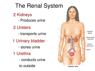
Introduction A urinalysis is a common test used to analyze the content and chemical makeup of your urine. While it is standardly performed before surgery to identify any kidney problems, a urinalysis may be used at a healthcare provider's office if a kidney infection, urinary tract infection, or other urinary-related disorder is suspected.
Purpose of Test Help diagnose medical conditions such as a urinary tract infection (UTI), kidney stones, uncontrolled diabetes, chronic kidney disease (CKD), acute renal failure, polycystic kidney disease (PKD), and kidney inflammation (glomerulonephritis) Screen for diseases such as kidney disease, diabetes, high blood pressure (hypertension), liver disease, and other conditions in which the urinary tract is involved
Purpose of Test Monitor disease progression and your response to treatment for kidney failure, diabetic nephropathy, lupus nephritis, and hypertension- related renal impairment, among others Provide a preoperative assessment of your renal function prior to undergoing surgery2 Monitor for pregnancy abnormalities, including bladder or kidney infection, dehydration, preeclampsia, and gestational diabetes, among others
Collection of urine Early morning sample-qualitative Random sample- routine 24hrs sample- quantitative Midstream sample-UTI Post prandial sample-D.M
24 hour urine sample 1. For quantitative estimation of proteins 2. For estimation of vanillyl mandelic acid, 5- hydroxyindole acetic acid, metanephrines 3. For detection of AFB in urine 4. For detection of microalbuminuria
Urine examination Macroscopic examination Chemical examination Microscopic examination
Macroscopic examination Volume Color Odour Reaction or urinary pH Specific gravity
Urinary volume Normal = 600-1550ml Polyuria- >2000ml Oliguria-<400ml Anuria-complete cessation of urine(<200ml) Nocturia-excretion of urine by a adult of >500ml with a specific gravity of <1.018 at night (characteristic of chronic glomerulonephritis)
Causes of polyuria Diabetes mellitus Diabetes insipidus Polycystic kidney Chronic renal failure Diuretics Intravenous saline/glucose
oliguria Dehydration-vomiting, diarrhoea, excessive sweating Renal ischemia Acute tubular necrosis Obstruction to the urinary tract Acute renal failure
Color & appearance 1. Colourless- dilution, diabetes mellitus, diabetes insipidus, diuretics 2. Milky-purulent genitourinary tract infection, chyluria 3. Orange-fever, excessive sweating 4. Red-beetroot ingestion,haematuria 5. Brown/ black- alkaptunuria, melanin Normal= clear & pale yellow
Urinary pH/ reaction Reaction reflects ability of kidney to maintain normal hydrogen ion concentration in plasma & ECF Normal= 4.6-8 Tested by- 1.litmus paper 2. pH paper 3. dipsticks
Acidic urine Ketosis-diabetes, starvation, fever Systemic acidosis UTI- E.coli Acidification therapy
Alkaline urine Strict vegetarian Systemic alkalosis UTI- Proteus Alkalization therapy
Odour Normal= aromatic due to the volatile fatty acids Ammonical bacterial action Fruity- ketonuria
Specific gravity Depends on the concentration of various solutes in the urine. Measured by-urinometer - refractometer - dipsticks
Urinometer Take 2/3 of urinometer container with urine Allow the urinometer to float into the urine Read the graduation at the lowest level of urinary meniscus Correction of temperature & albumin is a must. Urinometer is calibrated at 15or 200c So for every 3oc increase/decrease add/subtract 0.001 For 1gm/dl of albumin add0.001
High specific gravity(hyperosthenuria) Normal-1.016-1.022 Causes All causes of oliguria Gycosuria
Low specific gravity(hyposthenuria) All causes of polyuria except gycosuria Fixed specific gravity (isosthenuria)=1.010 Seen in chronic renal disease when kidney has lost the ability to concentrate or dilute
Chemical examination Proteins Sugars Ketone bodies Bilirubin Bile salts Urobilinogen Blood
Tests for proteins Test HEAT & ACETIC ACID TEST Principle-proteins are denatured & coagulated on heating to give white cloud precipitate. Method-take 2/3 of test tube with urine, heat only the upper part keeping lower part as control. Presence of phosphates, carbonates, proteins gives a white cloud formation. Add acetic acid 1-2 drops, if the cloud persists it indicates it is protein(acetic acid dissolves the carbonates/phosphates)
Other tests Sulphosalicylic acid test Dipsticks Esbach s albuminometer- for quantitative estimation of proteins
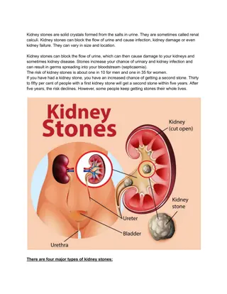
Causes of proteinuria Prerenal causes-Heavy exercise,Fever,hypertension, multiple myeloma, ecalmpsia Renal acute & chronic glomerulonephritis,Renal tubular dysfunction,Polycystic kidney, nephrotic syndrome Post renal- acute & chronic cystitis, tuberculosis cystitis
Selective proteinuria Nonselective proteinuria
microalbuminuria The level of albumin protein produced by microalbuminuria cannot be detected by urine dipstick methods. In a properly functioning body, albumin is not normally present in urine because it is retained in the bloodstream by the kidneys. Microalbuminuria is diagnosed either from a 24-hour urine collection
Significance of microalbuminuria an indicator of subclinical cardiovascular disease an important prognostic marker for kidney disease in diabetes mellitus in hypertension increasing microalbuminuria during the first 48 hours after admission to an intensive care unit predicts elevated risk for acute respiratory failure , multiple organ failure , and overall mortality
Bence Jones proteins These are light chain globulins seen in multiple myeloma, macroglobulimias, lymphoma. Test- Thermal method(waterbath): Proteins has unusual property of precipitating at 400 -600c & then dissolving when the urine is brought to boiling(1000c) & reappears when the urine is cooled.
Test for sugar Test-BENEDICT S TEST(semiquantitative) Principle-benedict s reagent contains cuso4.In the presence of reducing sugars cupric ions are converted to cuprous oxide which is hastened by heating, to give the color. Method- take 5ml of benedict s reagent in a test tube, add 8drops of urine. Boil the mixture. Blue-green= negative Yellow-green=+(<0.5%) Greenish yellow=++(0.5-1%) Yellow=+++(1-2%) Brick red=++++(>2%)
Benedicts test Detects all reducing substances like glucose, fructose, & other reducing sustances. To confirm it is glucose, dipsticks can be used (glucose oxidase)
Causes of glycosuria Glycosuria with hyperglycaemia- diabetes,acromegaly, cushing s disease, hyperthyroidism, drugs like corticosteroids. Glycosuria without hyperglycaemia- renal tubular dysfunction
Ketone bodies 3 types Acetone Acetoacetic acid -hydroxy butyric acid They are products of fat metabolism
Rotheras test Principle-acetone & acetoacetic acid react with sodium nitroprusside in the presence of alkali to produce purple colour. Method- take 5ml of urine in a test tube & saturate it with ammonium sulphate. Then add one crystal of sodium nitroprusside. Then gently add 0.5ml of liquor ammonia along the sides of the test tube. Change in colour indicates + test
Causes of ketonuria Diabetes Non-diabetic causes- high fever, starvation, severe vomiting/diarrhoea
Bilirubin Test- fouchet s test. Causes Liver diseases-injury,hepatitis Obstruction to biliary tract
Urobilinogen Test- ehrlich test Causes-hemolytic anemia's Bile salts- Hay s test Cause- obstruction to bile flow (obstructive jaundice)
Blood in urine Test- BENZIDINE TEST Principle-The peroxidase activity of hemoglobin decomposes hydrogen peroxide releasing nascent oxygen which in turn oxidizes benzidine to give blue color. Method- mix 2ml of benzidine solution with 2ml of hydrogen peroxide in a test tube. Take 2ml of urine & add 2ml of above mixture. A blue color indicates + reaction.
Causes of hematuria Pre renal- bleeding diathesis, hemoglobinopathies, malignant hypertension. Renal- trauma, calculi, acute & chronic glomerulonephritis, renal TB, renal tumors Post renal severe UTI, calculi, trauma, tumors of urinary tract
Type Plasma color Urine color Hematuria normal Smoky red m/s-plenty of RBC s Red , occasional RBC s Red, occasional RBC s hemoglobunuria Pink,hepatoglob in reduced Myoglobunuria Pink, normal hepatoglobin
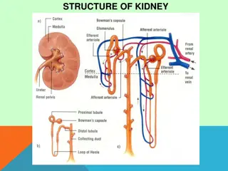
Microscopic examination Microscopic urinalysis is done simply pouring the urine sample into a test tube and centrifuging it (spinning it down in a machine) for a few minutes. The top liquid part (the supernatant) is discarded. The solid part left in the bottom of the test tube (the urine sediment) is mixed with the remaining drop of urine in the test tube and one drop is analyzed under a microscope
Contents of normal urine m/s Contains few epithelial cells, occasional RBC s, few crystals.
Crystals in urine Crystals in acidic urine Uric acid Calcium oxalate Cystine Leucine Crystals in alkaline urine Ammonium magnesium phosphates(triple phosphate crystals) Calcium carbonate
casts Urinary casts are cylindrical aggregations of particles that form in the distal nephron, dislodge, and pass into the urine. In urinalysis they indicate kidney disease. They form via precipitation of Tamm-Horsfall mucoprotein which is secreted by renal tubule cells.
Types of casts Acellular casts Hyaline casts Granular casts Waxy casts Fatty casts Pigment casts Crystal casts Cellular casts Red cell casts White cell casts Epithelial cell cast
Hyaline casts The most common type of cast, hyaline casts are solidified Tamm-Horsfall mucoprotein secreted from the tubular epithelial cells Seen in fever, strenuous exercise, damage to the glomerular capillary


")
")