Urinary Tract Infections in Children: Prevalence, Manifestations, and Management

Urinary tract infections (UTIs) are common in children, with different prevalence and risk factors based on age and gender. Learn about the clinical manifestations, causative agents, and management of UTIs in infants and children. Understand the types of UTIs like pyelonephritis and cystitis, and their impact on renal health. Discover the pathogenesis of UTIs and the diagnostic criteria for identifying these infections through urinalysis and culture.

Download Presentation

Please find below an Image/Link to download the presentation.
The content on the website is provided AS IS for your information and personal use only. It may not be sold, licensed, or shared on other websites without obtaining consent from the author.If you encounter any issues during the download, it is possible that the publisher has removed the file from their server.
You are allowed to download the files provided on this website for personal or commercial use, subject to the condition that they are used lawfully. All files are the property of their respective owners.
The content on the website is provided AS IS for your information and personal use only. It may not be sold, licensed, or shared on other websites without obtaining consent from the author.
E N D
Presentation Transcript
Urinary tract infection Dr. Nihal Khalid Yaseen Dr. Nihal Khalid Yaseen
1- Understand prevalence, risk factors, and causative agents for UTI in infants and children. 2- Identify clinical manifestations and management of UTI in infants and children. 3- Illustrate different types of VUR in children with their management. Learning objectives
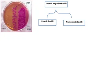
Background Urinary tract infections (UTIs) are common in children of all ages. During the first 3 months of life, UTIs are most common in uncircumcised febrile males with a prevalence of 20%. After 6 months of age, UTIs are much more likely in females, with peaks during infancy, toilet training, and onset of sexual activity. UTIs are caused primarily by colonic bacteria mainly Escherichia coli, followed by Klebsiella spp. and Proteus spp., Enterococcus, and Pseudomonas. Other bacteria known to cause UTIs include Staphylococcus saprophyticus, group B streptococcus, and, less commonly, Staphylococcus aureus, Candida spp., and Salmonella spp. Two basic forms of UTIs identified (pyelonephritis and cystitis). Focal pyelonephritis (lobar nephronia) and renal abscesses are less common.
Clinical manifestations Pyelonephritis (infection of the renal parenchyma) is characterized by: abdominal, or flank pain; fever; malaise; nausea; vomiting; and, occasionally, diarrhea. Fever may be the only manifestation, especially in young children; Newborns can also show nonspecific symptoms, such as poor feeding, irritability, jaundice, or weight loss. Pyelonephritis is the most common bacterial infection in infants younger than 24 months of age who have fever without an obvious focus. Cystitis (bladder infection) symptoms include dysuria, urgency, frequency, suprapubic pain, incontinence, and possibly malodorous urine. Cystitis does not cause high fever and does not result in renal injury. Malodorous urine is suggestive but not specific for a UTI. Acute hemorrhagic cystitis has been attributed to E. Coli, adenovirus types 11 and 2,or as side effect of chemotherapeutic agents. It is self- limiting, with hematuria lasting approximately 4 days.
Pathogenesis Nearly all UTIs are ascending infections. The fecal flora, colonize the perineum, and enter the bladder via the urethra. In uncircumcised males, the bacterial pathogens arise from the flora beneath the prepuce. In some cases, the bacteria causing cystitis ascend to the kidney to cause pyelonephritis. Rarely, renal infection occurs by hematogenous endocarditis or in bacteremic neonates. If bacteria ascend from the bladder to the kidney, acute pyelonephritis can occur. Infected urine stimulates an immunologic and inflammatory response that can cause renal injury and scarring. spread, as in
Diagnosis Diagnosis 1-Urinalysis findings suggestive of infection include:- pyuria > 5 WBC/mm3, positive leukocyte esterase, presence of nitrites, microscopic hematuria (in cystitis), and WBC casts. 2-Urine culture with 50,000 colony- forming units (CFU)/ mL of a single uropathogen in a symptomatic child. 3- Complete blood count shows leukocytosis and neutrophilia 4- Elevated ESR, procalcitonin level, and CRP. 5- Bacteremia occur in 3 20% of patients and is most common in infants.
Collection of a urine sample In toilet- trained children, a midstream urine sample usually is satisfactory; the introitus should be cleaned before obtaining the specimen. In uncircumcised males, the prepuce must be retracted. For children who are not toilet trained, a catheterized urine sample should be obtained. Alternatively, the application of an adhesive, sealed, sterile collection bag after disinfection of the skin of the genitals can be useful. A bagged specimen should not be the method because of a high rate of contamination. A suprapubic aspirate is unnecessary.
Sterile pyuria (positive leukocytes, negative culture) may occur in:- 1-partially treated bacterial UTI. 2-viral infections. 3-urolithiasis. 4-renal tuberculosis. 5-renal abscess. 6-sexually transmitted infection. 7-appendicitis, Crohn disease. 8-Kawasaki disease. 9-schistosomiasis. 10-interstitial nephritis.
Imaging The primary goal of imaging studies in children with a febrile UTI is to identify anatomic abnormalities that predispose to future infection, such as high- grade VUR, posterior urethral valves, or other obstructive uropathy. 1- Ultrasound is the first- line imaging for screening recommended for all children with UTI and will likely demonstrate an enlarged kidney with a possible mass in the case of acute lobar nephronia or renal abscess. 2- CT scan is more sensitive and specific for lobar nephronia and will typically show a wedge- shaped, lower- density area after contrast administration. 3- voiding cystourethrogram (VCUG) done only only if atypical features (including non E. coli infection, sepsis or bacteremia, failure to respond to antibiotics within 48 hours), recurrent UTI, or abnormal ultrasound findings. 4- dimercaptosuccinic acid (DMSA) renal scan.
Treatment 1- Oral treatment, used in most patients as an outpatient setting, oral cephalexin is a commonly used narrow- spectrum empiric option, also oral third- generation cephalosporins such as cefixime, nitrofurantoin, Trimethoprim- sulfamethoxazole (TMP- SMX), and oral fluoroquinolone such as ciprofloxacin are increasingly used. 2- Parenteral therapy should be used in children who are dehydrated, are vomiting, are unable to drink fluids, have complicated infection, or in whom urosepsis is a possibility. Infants less than 1 month of age with suspected febrile UTI are typically hospitalized and started on parenteral antibiotics while awaiting results of a sepsis evaluation. ceftriaxone, ampicillin plus either gentamicin or a third- generation cephalosporin are treatment options. 3- 3- Acute lobar nephronia, renal abscess, and perinephric abscess usually treated with the same antibiotics as pyelonephritis. The recommended duration of treatment is 14- 21 days in addition to percutaneous drainage if necessary.
Prevention Prevention In a child with recurrent UTIs, identification of predisposing fac tors is beneficial. Behavioral modification, with treatment of constipation is effective at reducing recurrent UTI from constipation. Routine use of antibiotic prophylaxis is not recommended for children with a first episode of pyelonephritis and an otherwise anatomically normal urinary tract. Urologic conditions that can cause recurrent UTIs that might benefit from long- term antibiotic prophylaxis include neurogenic bladder, urinary tract stasis and obstruction, severe VUR, and urinary calculi. Prophylactic agents as: - Trimethoprim or nitrofurantoin at 30% of the normal therapeutic dose once a day. Amoxicillin calvulunate, or cephalexin can also be effective
Vesico ureteral reflux: Defined as retrograde flow from the bladder to the ureter and kidney. Affecting 1 2% of children and is usually congenital and familial. VUR predisposes to pyelonephritis by facilitating the transport of bacteria from the bladder to the upper urinary tract. The inflammatory reaction caused by pyelonephritis can result in renal injury or scarring, also termed reflux-related renal injury or reflux nephropathy. Scarring associated with reflux may be present at birth or develop in the absence of infection if there is significant bladder- sphincter discoordination during voiding.
Classification Grade I: VUR into a nondilated ureter. Grade II: VUR into the upper collecting system without dilation. Grade III: VUR into a dilated ureter and/or blunting of the calyceal fornices. Grade IV: VUR into a grossly dilated ureter. Grade V: massive VUR, with significant ureteral dilation and tortuosity and loss of the papillary impression.
Clinical manifestations Clinical manifestations VUR usually is discovered during evaluation for a UTI, antenatal hydronephrosis, or chronic kidney disease. The UTI may be symptomatic, an isolated febrile event, or more often both febrile and symptomatic. Constipation may be present in 50% of children with reflux and a UTI.
Management VCUG or radionuclide cystogram is done for diagnosis. Grade I & II who are toilet trained offered watchful waiting while those who are not toilet trained need antibiotic prophylaxis. Grade III & IV are managed by prophylactic antibiotics with surveillance for infection. Grade V need surgery (ureteric reimplantation).
References Nelson textbook f pediatric, 22nd edition, 2024 UpToDate


 may")