Using Vital Dyes for Ophthalmic Disease Diagnosis
Vital dyes play a crucial role in diagnosing and evaluating ocular surface disorders, with properties like water solubility, selective cell staining, and compatibility with eye procedures. Fluorescein and Rose Bengal are commonly used dyes with specific indications, routes of administration, and precautions to consider for safe usage.

Download Presentation

Please find below an Image/Link to download the presentation.
The content on the website is provided AS IS for your information and personal use only. It may not be sold, licensed, or shared on other websites without obtaining consent from the author.If you encounter any issues during the download, it is possible that the publisher has removed the file from their server.
You are allowed to download the files provided on this website for personal or commercial use, subject to the condition that they are used lawfully. All files are the property of their respective owners.
The content on the website is provided AS IS for your information and personal use only. It may not be sold, licensed, or shared on other websites without obtaining consent from the author.
E N D
Presentation Transcript
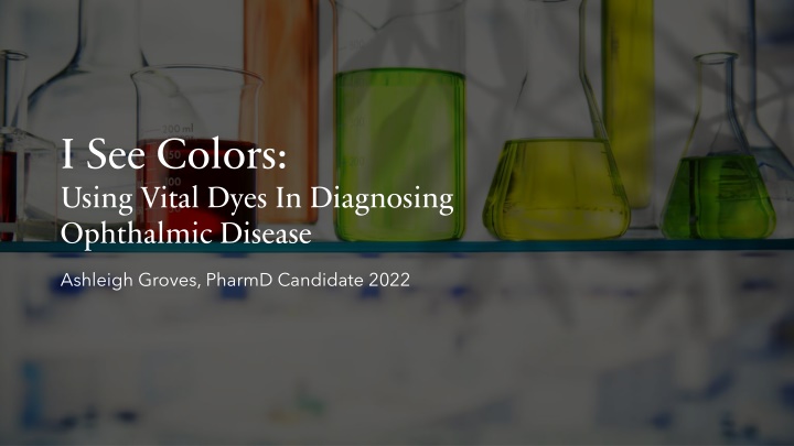
I See Colors: Using Vital Dyes In Diagnosing Ophthalmic Disease Ashleigh Groves, PharmD Candidate 2022
Vital Dyes Utilized for diagnosing and evaluating ocular surface disorders Commercially available vs. compounded products Preservative free formulations Availability in the U.S.
Ideal Properties Water Soluble Selectively stain cells or structures Should not stain skin, clothing, contacts, or instruments Reversible effect No vision interference No pharmacologic effects Non-irritant and non-toxic to the eye Compatible with other stains and agents used in the eye
Fluorescein What: Orange-red dye that fluoresces in high dilution Purpose: Detects epithelial defects and assesses healing of corneal epithelial lesions Indications: Diagnostic aid in angiography or angioscopy of the retina and iris vasculature To stain the anterior segment of the eye for procedures (contact lens fitting), disclosing corneal injury, and in applanation tonometry Assessment of tear break-up time (BUT); tear flow assessment Determination of nasolacrimal duct patency
Fluorescein Route of Administration: IV or ophthalmic strips IV: 500 mg as single dose into antecubital vein Ophthalmic strips: Moisten strip with sterile water, saline, or ophthalmic fluid. Touch conjunctiva or fornix with tip of strip until adequately stained. Instruct patient to blink frequently. Caution: Utilize test dose in individuals with suspected allergy Warnings: Increased risk of microbial contamination (esp. Pseudomonas aeruginosa) with ophthalmic solution Include preservative phenylmercuric acetate or nitrate 0.002% Use sterile single-dose units
Rose Bengal What: Brownish-red powder that turns dark magenta when dissolved in water as a 1% solution Purpose: Stains dead cells and mucus threads Indications: Dendritic keratitis to stain the ulcer Keratoconjunctivitis sicca (dry eye) Keratitis neuroparalytica Exophthalmos Pressure areas due to contact lens wear Delineation the extent of corneal and conjunctival neoplasms
Rose Bengal Route of Administration: IV or ophthalmic strips Warnings: Irritation Pretreat patient with a topical anesthetic Photothrombosis when combined with arterial irradiation Antiviral effects
Lissamine Green B What: Similar characteristics to Rose Bengal Purpose: Stains lipid-like structures, dead or dying epithelial cells, and leading edges of viral proliferation of a dendritic ulcer Advantages over Rose Bengal: Does not possess antiviral activity Not as irritating/better tolerated Equally effective
Mixed Products Provide more comprehensive exam Fluorescein 1% + Rose Bengal 1% Fluorescein preferred in corneal staining Rose Bengal preferred in bulbar conjunctival staining Uncomfortable to patient Fluorescein 1% + Lissamine Green B 1% Better tolerated by patients Lissamine has replaced the use of Rose Bengal Reprinted from Contact Lens Complications3
Brilliant Blue G Purpose: Used in macular surgery to visualize the internal limiting membrane (ILM) Indication: ILM stain Route of Administration: Intravitreal Intravitreal: Inject 0.5 mL into a balanced salt solution-filled vitreous cavity
Brilliant Blue G Comparable products: Indocyanine green (ICG) & Trypan blue (TB) Advantage: Does not require active aspiration to remove the dye, unlike ICG and TB Superior to ICG and TB Must be compounded as a high-risk sterile preparation
Additional Dyes Dyes Alcian blue Uses Counterstain Rose Bengal, stains dead cells and mucus Stains mucus and dead cells which have undergone structural changes; ILM staining Staining of tumors and assessing corneal grafts Used to assess damage caused by chemical agents such as lime Bacterial stain that also vitally stains nerve tissue; stains ulcers ILM staining Trypan blue Tetrazolim and iodonitrotetrazolium Bromothymol blue Methylene blue Indocyanine green
References McElhiney, Linda F., PharmD, RPh, FIACP, FASHP, FACA, I See Colors: Using Vital Dyes in Diagnosing Ophthalmic Disease. International Journal of Pharmaceutical Compounding. 2012; 16(3): 190-195. Fluorescein. Lexi-Drugs. Lexicomp. Wolters Kluwer. Hudson, Oh. Available at https://online.lexi.com. Accessed May 11, 2021. Nathan Efron, Chapter 16 - Corneal Staining, Editor: Nathan Efron, Contact Lens Complications (Third Edition), W. B. Saunders, 2012, Pages 155-156, ISBN 9780702042690, https://doi.org/10.1016/B978-0- 7020-4269-0.00016-X. Accessed May 11, 2021. Brilliant Blue G. Lexi-Drugs. Lexicomp. Wolters Kluwer. Hudson, Oh. Available at https://online.lexi.com. Accessed May 11, 2021.