Uterine Leiomyomata: Etiology, Pathology, and Growth
Uterine leiomyomata, benign tumors mainly of smooth muscle cells, have varying growth characteristics and are common in women. Factors influencing growth include estrogen production, postmenopausal changes, and individual risk factors like parity, smoking, and obesity.

Download Presentation

Please find below an Image/Link to download the presentation.
The content on the website is provided AS IS for your information and personal use only. It may not be sold, licensed, or shared on other websites without obtaining consent from the author.If you encounter any issues during the download, it is possible that the publisher has removed the file from their server.
You are allowed to download the files provided on this website for personal or commercial use, subject to the condition that they are used lawfully. All files are the property of their respective owners.
The content on the website is provided AS IS for your information and personal use only. It may not be sold, licensed, or shared on other websites without obtaining consent from the author.
E N D
Presentation Transcript
LEIOMYOMATA UTERI AND MYOMECTOMY
ETIOLOGY, PATHOLOGY, AND GROWTH CHARACTERISTICS OF UTERINE LEIOMYOMATA A leiomyoma is a benign tumor composed mainly of smooth muscle cells but containing varying amounts of fibrous connective tissue The tumor is well circumscribed but not encapsulated Leiomyomata are the most common tumors of the uterus and female pelvis Leiomyomata are responsible for about one third of all hospital admissions to gynecology services It is well recognized that the incidence is much higher in African American than in Caucasian women.
ETIOLOGY, PATHOLOGY, AND GROWTH CHARACTERISTICS OF UTERINE LEIOMYOMATA Patients with uterine leiomyomata often have a positive family history of uterine leiomyomata. About 40% to 50% of leiomyomas show karyotypically detectable chromosomal abnormalities: Identified chromosomal abnormalities include t(12;14) (q15;q23-24), del(7) (q22q32), rearrangements involving 6p21, 10q, trisomy 12, and deletions of 3q.
The growth of leiomyomata is dependent on estrogen production. After menopause, with regression of ovarian estrogen secretion, growth of leiomyomata usually ceases. However, of postmenopausal growth of benign leiomyomata, suggesting the possibility of postmenopausal estrogen production either in the ovary or elsewhere.
Postmenopausal ovarian cortical stromal hyperplasia may be associated with an increase in estrogen secretion by the ovary. The postmenopausal ovarian stroma in a variety of presumably inactive ovarian tumors, including mucinous cysts and Brenner tumors, can also produce estrogen. When a central pelvic tumor presumed to beuterine leiomyomata enlarges after menopause, one should think of the possibility of malignant change in the leiomyoma itself or in the adjacent myometrium, or of the growth of a new pelvic tumor of extrauterine origin.
Older nulliparous women have an increased risk of developing leiomyomata. However, in multiparous women, the relative risk decreases with each pregnancy. The risk is reduced in women who smoke and is increased in obese women; this is possibly related to the conversion of androgens to estrogen by fat aromatase.
The observation that leiomyomata may show significant enlargement during pregnancy provides further clinical evidence of the relation of estrogen and progesterone to the growth of these tumors. However, a better blood supply during pregnancy might also encourage their growth.
Oral contraceptives containing high-dose estrogen, there was a striking increase in the occurrence of large leiomyomata Oral contraceptives containing highdose estrogen should not be prescribed for women with these tumors. Oral contraceptives with low-dose estrogen are less likely to stimulate growth.
Levels of plasma estradiol were the same in patients with and without leiomyomata. Significantly higher concentration of estrogen receptors in leiomyomata than in myometrium. The enzyme 17 -hydroxy dehydrogenase accelerates the conversion of estradiol to estrone. Leiomyomata low concentration of 17 - hydroxy dehydrogenase Hypertrophy Myometrial
Abnormal pituitary function in women with leiomyomata: low FSH level and a diminished FSH response to pituitary GnRH. There was an excessive prolactin response to TRH. Peak levels of human growth hormone during a hypoglycemic test o Significant increase in 5 -reductase
Both estrogen and progesterone are involved in the growth of uterine leiomyomata. Polypeptide growth factors that have been investigated include epidermal growth factor, transforming growth factor alpha and beta, insulin-like growth factor (IGF), platelet-derived growth factor, vascular endothelial growth factor, and basic fibroblast growth factor.
Leiomyomata may be single, but most are multiple. They develop most commonly in the uterine corpus and much less often in the cervix. They may develop in the round ligaments, but this is rare. Because they arise in the myometrium, they are all interstitial or intramural in the beginning. Subserous or Submucous
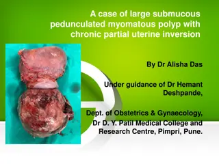
A subserous tumor can become pedunculated and occasionally parasitic, receiving its blood supply from another source, usually the omentum. A submucous tumor can also become pedunculated and may gradually dilate the endocervical canal and protrude through the cervical os. Indeed, a submucous myoma may descend through the vagina. Rarely, chronic uterine inversion results if the prolapsing submucous leiomyoma is attached to the top of the endometrial cavity and pulls the uterine fundus downward through the cervix.
Subserous and subserous pedunculated tumors, as well as intraligamentous tumors, may create problems in diagnosis because they are difficult to distinguish from tumors arising from the adnexal organs Ordinarily, there is a clear distinction between the myoma and the myometrium so that dissection between the two is easy to accomplish Although these tumors are not encapsulated, a clear distinction can usually be made between a myoma and the myometrium that surrounds it. The cut surface appears as glistening pinkish white and gray
HYALINE DEGENERATION The most common change The cut surface of a hyalinized area is smooth and homogeneous These may become liquefied and form cystic cavities filled with clear liquid or gelatinous material
CALCIFIED LEIOMYOMATA Over time, with continued diminished blood supply and ischemic necrosis of tissue, calcium phosphates and carbonates are deposited in myomata. When the degenerative change is advanced, the leiomyoma may become solidly calcified. Such calcified tumors have been called womb stones. Most often in elderly women, in African American women, and in women who have pedunculated subserous tumors. They are easily seen radiographically
INFECTION Submucous leiomyomata are most commonly infected when they protrude into the uterine cavity, or especially into the vagina An intramural leiomyoma in an involuting puerperal uterus can also become infected when endometritis is present. Such infections are usually Streptococcal and may be virulent. Bacteroides fragilis infections also occur. Parametritis, peritonitis, and even septicemia may result.
Necrosis of a leiomyoma is caused by interference with its blood supply. Occasionally, a pedunculated subserous leiomyoma twists, and if an operation is not done immediately, infarction results.
RED OR CARNEOUS DEGENERATION Necrotic leiomyomata are dark and hemorrhagic in the interior. Eventually, the tissue breaks down completely. So-called red or carneous degeneration is seen occasionally, especially in association with pregnancy. This condition is thought to result from poor circulation of blood through a rapidly growing tumor. Thrombosis and extravasation of blood into the myoma tissue are responsible for the reddish discoloration
FATTY DEGENERATION Fat occurs in leiomyomata as true fatty degeneration The cut surface may have a yellowish discoloration. Infrequently, a deposit of true fat may form a fibrolipoma; however, the presence of fat in a leiomyoma is rare. If fat is seen grossly or microscopically in a curettage specimen, one should not assume that it represents fatty degeneration of a leiomyoma. One should assume that the uterus has been perforated and that fragments of fat have been curetted from the mesentery or omentum.
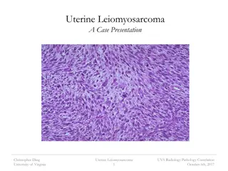
SARCOMATOUS DEGENERATION The most important, but rare, change in a leiomyoma A sarcoma is likely to occur in a rather large leiomyoma and toward the center of the tumor, where the blood supply is poorest. The tissue is soft and homogeneous and is described as resembling raw pork. Later, as necrosis of the malignant tissue occurs, it becomes more friable and hemorrhagic.
All tumors with less than 5 mitotic figures per 10 high-power fields are considered benign. All tumors with more than 10 mitotic figures per 10 high-power fields are called malignant. Those in between can be called smooth muscle tumors of uncertain malignant potential. Presence of nuclear hyperchromatism, nuclear pleomorphism, or giant cells and other bizarre cell forms to makethe diagnosis. Smooth muscle tumors that metastasize or recur are definitely malignant
A normal chromosome complement (46,XX) About 50% showed clonal abnormalities, such as those of chromosomes 1, 7, and 13, and t(12;14).
INTRAVENOUS LEIOMYOMATOSIS The characteristic feature of this peculiar smooth muscle tumor is the extension of the polypoid intravascular projections into the veins of the parametrium and broad ligaments. The uterine veins in the broad ligaments are the most common sites of extension clinically like a benign neoplasm Total surgical excision of the tumor should be attempted for successful therapy Extension of benign leiomyomatosis up the vena cava and into the right atrium has been reported
Several recent cases requiring open heart surgery to remove the intracardiac tumor thrombosis have been successful and without recurrence Both intravenous leiomyomatosis and benign metastasizing leiomyoma have been reported to metastasize to the lung Oophorectomy may be indicated in patients with these conditions
LEIOMYOMATOSIS PERITONEALIS DISSEMINATA Only subperitoneal surfaces of the uterus and other pelvic and abdominal viscera are involved Invasion of the lumen of blood vessels does not occur. All occurred in patients in the reproductive years who often had large uterine leiomyomata and were usually pregnant or taking oral contraceptives The tumor is benign, and the acceptable treatment to date is total abdominal hysterectomy and bilateral salpingo-oophorectomy If this tumor occurs in the omentum, an omentectomy should also be performed to define more clearly the histologic nature of the lesion.
VASCULARIZATION OF LEIOMYOMATA Leiomyomata have a rich vascular supply, including blood lakes within tumors. Venous channels were predominantly peripheral, whereas the arterial supply was both internal and peripheral.
CLINICAL FEATURES OF UTERINE LEIOMYOMATA Most leiomyomata are asymptomatic Small leiomyomata that are asymptomatic need only to be observed from time to time, with pelvic examinations perhaps every 6 to 12 months and pelvic ultrasonography (US) when indicated. If small leiomyomata are discovered late in menstrual life, it is unusual for symptoms to appear or for surgical treatment to be required.
surgical removal of leiomyomata when the uterus reaches 12 weeks gestational size or greater, regardless of the presence or absence of significant symptoms. The inability to accurately assess the ovaries by examination The possible malignancy of the pelvic mass The potential for compromise of adjacent organ function if the mass continues to enlarge The greater risk of surgical complications if the mass grows to a larger size The potential for better fertility if myomectomy is performed when the uterus is smaller The possibility of continued growth of uterine leiomyomata if hormone replacement therapy is given after menopause
If one elects to observe a patient with a relatively large asymptomatic uterine leiomyoma, it is a good rule to obtain an excretory urogram or renal ultrasound. Ureteral compression at the pelvic brim may occur so that hydroureter and hydronephrosis develop
In the postmenopausal years, shrinkage of myomata and the myometrium occurs. However, the myometrial shrinkage may be disproportionately greater than the myoma shrinkage. Therefore, a myoma in an intramural location before menopause may become a submucous myoma after menopause and then become symptomatic for the first time, usually with postmenopausal bleeding. In menopausal women, the appearance of even the slightest trace of vaginal bleeding should make one suspect cervical or endometrial malignancy or the possibility of sarcomatous change in the leiomyoma
DETECTION OF LEIOMYOMAS Transabdominal and endovaginal US Sonohysterography CA-125 levels may be elevated in women with uterine leiomyomata, but the levels are generally lower than those in patients with ovarian cancer.
SYMPTOMATIC LEIOMYOMATA Abnormal Bleeding Pressure Pain Abdominal Distortion Rapid Growth:a gain of 6 weeks or more in gestational size within a year or less Spontaneous Abortion and Other Pregnancy- Related Problems Infertility Miscellaneous Signs and Symptoms
CHOICE OF TREATMENT FOR UTERINE LEIOMYOMATA Medical Management of Uterine Leiomyomata Effective medical treatment that is likely to result in the permanent cure of uterine leiomyomata is not yet available. Medical therapies are available as an adjunct to surgical treatment or as a temporary substitute for definitive surgical treatment.
Antiprogestin therapy with mifepristone (RU-486) 25 mg daily decrease leiomyoma volume by an average of 49% for 3 months All patients became amenorrheic Side effects were mild, and bone density was not diminished o Gestrinone antiestrogen and antiprogesterone properties The treatment lasted 6 months to 1 year. The best results were obtained when the drug was administered intravaginally Side effects, though mildly androgenic, were well tolerated
GnRH analogs medical oophorectomy and medical menopause describe a significant reduction in trabecular bone density after 24 weeks of GnRH agonist treatment the average reduction in uterine and myoma volume is 40% to 50% after 3 to 6 months of GnRH agonist treatment If there are circumstances that require that GnRH treatment be extended beyond 6 months, consideration should be given to adding low-dose steroids after 3 months of GnRH therapy
Hysteroscopic Resection of Submucous Myomata Laparoscopic and Robotic-Assisted Myomectomy Abdominal Myomectomy
Position the patient in Allen stirrups. Perform exam under anesthesia, paying careful attention to uterine size and mobility. Prep and drape the patient. Place Foley. Perform cervical dilation, and consider staining the endometrial cavity. Make an abdominal incision (consider Maylard for uteri >12-week size), and enter the abdominal cavity. Optimize exposure (incision type and size, lighting, packing of intestines, retractors). Evaluate number, size, and location of fibroids.
Minimize blood loss. Make use of delicate surgical technique (avoid traumatic instruments). Consider hypotensive anesthesia. Consider use of a tourniquet. Consider use of dilute Pitressin. Make a linear uterine incision over the largest myoma. Dissect along the natural tissue plane, and excise the myoma. Meticulously close all uterine defects. Confirm hemostasis. Close abdominal cavity, fascial, and skin incisions.
DECREASE SURGICAL BLEEDING Controlled hypotensive anesthesia Trendelenburgposition Tourniquets Vasoconstrictive agents The CO2 laser
RESULTS OF MYOMECTOMY The major immediate postoperative complications after myomectomy are febrile morbidity and intraperitoneal bleeding. patients must be carefully monitored for the first 24 hours after myomectomy A hematocrit is usually performed 6 hours after the operation is completed and again on the first postoperative morning
EMBOLOTHERAPY UAE is appropriate for patients with symptomatic leiomyomata Contraindications to this procedure include pregnancy, active pelvic infection, severe contrast medium allergy, arteriovenous malformations, desire for future pregnancy, a strong suspicion of adenomyosis or pedunculated leiomyoma, and undiagnosed pelvic mass. may increase the risk of miscarriage, postpartum hemorrhage, preterm delivery, and malpresentation
POSTEMBOLIZATION SYNDROME Forty percent of patients develop a syndrome of fever and malaise in the first 10 to 14 days after UAE. This is also associated with leukocytosis. It is typically selflimited and resolves in 3 to 5 days and rarely requires treatment except antipyretics.
UAE . . . .1 .2 .3 . .4


")