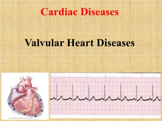
Valvular Heart Disease and Endocarditis
Valvular heart disease encompasses various conditions affecting the heart valves, including infectious and non-infectious endocarditis, valvular malformations such as stenosis and insufficiency. Learn about the clinical presentation, diagnostic methods, etiology, and pathology of infectious endocarditis in detail.

Download Presentation

Please find below an Image/Link to download the presentation.
The content on the website is provided AS IS for your information and personal use only. It may not be sold, licensed, or shared on other websites without obtaining consent from the author.If you encounter any issues during the download, it is possible that the publisher has removed the file from their server.
You are allowed to download the files provided on this website for personal or commercial use, subject to the condition that they are used lawfully. All files are the property of their respective owners.
The content on the website is provided AS IS for your information and personal use only. It may not be sold, licensed, or shared on other websites without obtaining consent from the author.
E N D
Presentation Transcript
Cardiovascular pathology Valvular heart disease
Plan General characteristic Diseases of endocardium - Infectious endocarditis - Non-infectious endocarditis Valvular malformations - Mitral and aortal stenosis - Mitral and aortal insufficiency - Clinical picture and diagnostics
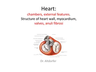
General characteristic Heart contains: Endocardium is distinguished to mural (wall) endocardium and to valvular endocardium Two atrioventricular valves Mitral Tricuspid Two semilunar valves Aortic Pulmonic
Infectious endocarditis Vegetation at valve > thrombus made from thrombocytes and fibrin If we observe microorganisms (MO) > infectious vegetation > infectious endocarditis In preATB era mostly young patients were affected by low virulent MO (alfa viridans streptococci) at rheumatic or malformed valves Now patients are older (50-60 y.o., 2:1 male to female), clinical picture is more pronounced and MO is more aggressive (golden staphylococci) at degenerative or after-surgery valves Mortality is still high (20-30%)
Infectious endocarditis. Etiopathogenesis The causative organisms - S. aureus, HACEK group (Haemophilus, Actinobacillus, Cardiobacterium, Eikenella, and Kingella) + mycotic infection Bacteremia is a MUST. Infection entrance can be anywhere surgeries, teeth extractions, catheterization . also narcomania Infection usually sits on changed valves (degenerative or iatrogenic) MO damages endothelium, thrombus formation starts, MO are multiplying inside thrombus, organism reacts with purulent inflammation
Infectious endocarditis. Pathology Progression of inflammation is often very fast Predilected places are atrial part of tricuspid and mitral valve, ventricular parts of pulmonar and aortic valves Left side valves are affected more commonly (mitral mostly, aortal less common) 10% of cases mitral and aortic valves are affected together In right sided IE tricuspid valve is always affected 5% of cases would be pulmonar, mural endocardium or zone of defect in ventricular wall Vegetation can be from some mm to 2cm
Clinical picture IE has been classified into acute and subacute forms. Acute infective endocarditis is typically caused by infection of a previously normal heart valve by a highly virulent organism that produces necrotizing, ulcerative, destructive lesions. Usually require surgery. Death within days to weeks. Subacute IE, the organisms are of lower virulence. These organisms cause insidious infections of deformed valves that are less destructive. Disease can last weeks to months, and cures are often produced with antibiotics.
Complication General extracardial Central pyemia Left-sided spread leads to abscess in brain tissue, purulent meningitis or haemorrhage, can spread to kidney or to coronary arteries Right-sided spread leads to purulent embols to lung artery Periferal microembolization subungual liner hematomas, Osler nodules, Roth plaques Immunocomplex disorder immunocomplex glomerulonephritis Local cardial Volume of vegetations can narrow valve, mix up with its closing or even damage it All of that can lead to acute heart failure Inflammation can spread locally and create ulcerations in endocardium and myocardial abscess
Special types of IE Endocarditis at valve transplant Endocarditis at intravenous drug addicts Nosocomial endocarditis
Non-bacterial (non-infection) endocarditis Noninfected (sterile) vegetations are caused by two mechanisms immunologic one (i.e. SLE) and hypercoagulation (thrombotic endocarditis)
Rheumatic endocarditis Chronic rheumatic disorder Late outcome of rheumatic fever Fibrous deformation of valves because of healing of fibrinoid necrosis with granulation tissue Leads to pronounced fibrotization and calcification of valves Acute rheumatic disorder 2/3 of cases Mostly pancarditis > serofibrinous inflammation, myocarditis with Aschoff nodules, endocarditis verrucosa (mitral mostly)
Endocarditis with SLE Appearence of immunocomplexes Inflammation happens in whole heart Typically it is lymphocytic myocarditis + non-bacterial endocarditis (Libman-Sacks) = sterile verrucous overgrowths mostly at left-side (mitral and aortal valve.)
Non-bacterial thrombotic endocarditis Cachectic patients marantic endocarditis Etiology is hypercoagulative condition which leads to thrombosis of endocardium. In 60% is a picture of paraneoplastic syndrome In 40% is a picture of chronic disease Mostly is found after 70 y.o. Vegetations are small 3-4 mm Mostly found at mitral valve
Valvular heart disease Types of valvular heart disease depend on Valve or valves affected Two types of alterations: Stenosis Insufficiency (regurgitation)
Definition Stenosis is the failure of a valve to open completely, which impedes forward flow. Insufficiency results from failure of a valve to close completely, thereby allowing reversed flow (regurgitation). These abnormalities can be present alone or coexist, and may involve only a single valve (isolated disease) or more than one valve (combined disease).
Pathophysiology Stenosis- narrowed valve, increases afterload Insufficiency- increases preload. The heart has to pump same blood All valvular diseases have a characteristic murmur
Valvular heart disease Valvular disorders occur: in children and adolescents primarily from congenital conditions in adults from degenerative heart disease Risk Factors Rheumatic Heart Disease Congenital Heart Defects Aging
Main reasons for acquired valvular disorders Aortal valve Mitral valve Aortic stenosis: Valvular diseases > - degenerative calcification - calcification of inherited valve malformation - rheumatic disorders Mitral stenosis: - Rheumatic disorder - Infectious endocarditis - Massive subanular calcification - Myxoma Aortic insufficiency: Valvular diseases > - Myxoid degeneration - Infection endocarditis - Rheumatic disorder - Bicuspidal aortal valve Abnormalities of aorta root Degenerative changes in aortal wall > - Aorta dissection - Systemic connective tissue diseases - Aortitis Mitral insufficiency: Disorders of leaflet > - Mitral prolapse myxoid degeneration - Rheumatic disorder - Infectious endocarditis Disorder of connective heart apparatus > - infarct/rupture/scarring of papillary muscles - Fibroelastic deficiency Disorders of form of LV or anulus of mitral valve > - Hypertrophy/dilatation of LV - Annular calcification
Aortic stenosis The most common age- associated wear and tear , most commonly with calcinosis On previously normal valves age 70-90 yo, bicuspid valves age 50- 70 yo heaped-up calcified masses within the aortic cusps that protrude through the outflow surfaces, preventing the opening of the cusps left ventricular myocardium to progressively increasing afterload
Aortic Stenosis Clinical Features - gradual narrowing of the valve orifice (up to 0.5 to 1 cm2; N 4 cm2) and an increasing pressure gradient. Left ventricular pressures rise, producing concentric left ventricular hypertrophy. - The hypertrophied myocardium ischemia angina pectoris. - Both systolic and diastolic myocardial function may be impaired.
Calcific Stenosis of Congenitally Bicuspid Aortic Valve the most frequent congenital cardiovascular malformation uncomplicated early in life, late complications - aortic stenosis or regurgitation, infective endocarditis, and aortic dilation and/or dissection Structural abnormalities of the aortic wall commonly accompany BAV there are only two functional cusps, usually of unequal size Valves can be changed congenitally or become bicuspid because of an acquired deformity (Rheumatism) clinical course is similar
Aortic insufficiency Less common Occurs in between 40-60 y.o. mostly at women Occurs in aorta root or in leaflets Leaflets are affected because of myxoid degeneration, rheumatic disorder, IE Aorta root is affected because of connective tissue disorder (Marphan syndrome) Part of blood volume of LV is returning in diastole through unclosing valve which leads to overload of LV Outcome is eccentric hypertrophy = cor bovinum (>1000 g)
Aortic insufficiency Clinical picture Exertional dyspnoea, angina pectoris, palpitations Long standing disease with good prognosis Calcification usually does not occur
Fish mouth Mitral stenosis 98% of cases of rheumatic etiology Usually occurs at old age, at really degenerated valves Stenosis leads to dilatation of LA, increased pressure goes to lung vessels where lung hypertense appears It increases pressure to RV which leads to systolic dysfunction and to dilatation of RA
Mitral insufficiency Can be primary (valve disorder) and secondary (remodulation of LV) The most common is mitral prolapse on the basis of myxoid valve degeneration Fibroelastic deficience is a form of pathologic ageing and degeneration Secondary mitral regurgitation is happening due to remodulation of LV
Mitral valve Normal open mitralvalve No Norm rmal closed al closed Prolapsed mitral va Prolapsed mitral valv lve; e; one leaflet does not one leaflet does not close properly close properly
Mitral Valve Prolapse Clinical picture May or may not be present with chest pain If pain occurs, episodes tend to occur in clusters, especially during stress Pain may be accompanied by dyspnea, palpitations, and syncope Does not respond to antianginal treatment
Less common valve disorders Pulmonary stenosis Pulmonary insufficience Tricuspidal stenosis Tricuspidal insufficience


 endocarditis")