Veterinary Medicine Insights: Viral Diseases of Zoo and Wild Animals
Explore common viral diseases affecting zoo and wild animals, including Foot and Mouth Disease (FMD) and Rabies. Learn about the etiology, epidemiology, clinical signs, diagnosis, and specimen collection methods for these diseases. Stay informed to protect animal health and welfare.

Download Presentation

Please find below an Image/Link to download the presentation.
The content on the website is provided AS IS for your information and personal use only. It may not be sold, licensed, or shared on other websites without obtaining consent from the author. If you encounter any issues during the download, it is possible that the publisher has removed the file from their server.
You are allowed to download the files provided on this website for personal or commercial use, subject to the condition that they are used lawfully. All files are the property of their respective owners.
The content on the website is provided AS IS for your information and personal use only. It may not be sold, licensed, or shared on other websites without obtaining consent from the author.
E N D
Presentation Transcript
Department of Veterinary Medicine Bihar Veterinary College, BASU, Patna Viral Diseases of Zoo and Wild animals Dr Mritunjay Kumar Associate Professor VMD, BVC, BASU, Patna
Common Viral Diseases of Zoo and Wild animals 1. FMD 2. Rabies 3. Canine Distemper 4. Avian influenza 5. Measles (Rubela) 6. Infectious Canine hepatitis 7. Kyasanur forest disease (KFD) 8. Herpesvirus virus infection 9. Feline Pan leukopenia 10. Parvo virus infections 11.Feline Infectious Peritonitis 12.Pox virus
Foot and Mouth Disease (FMD) Foot and Mouth Disease (FMD) Etiology Aphtha virus belonging to family Picornaviridae Positive sense single stranded RNA virus and is the smallest known virus of animal origin Seven major serotypes viz O, A, C, Asia 1 and SAT 1, SAT 2 and SAT 3 Epidemiology Affects all cloven footed artiodactyla Pigs including wild boars are main source of infection Unprocessed contaminated meat and meat products and contaminated feed, utensils may spread the virus Morbidity rate is very high and many animals affected same time Virus may survive for 1 year in infected premises
Clinical signs Fever, profuse salivation, vesicular lesions in udder, vesicles in mouth and feet, sudden death in young animals PM findings Vesicular erosion/ ulcerative stomatitis, oesophagitis Vesicle and erosion on feet and udder Tiger heart appearance in young animals Diagnosis Clinical signs and symptoms Virus isolation and typing and positive virus neutralization, ELISA, CFT, RT-PCR Specimen Collection Epithelial suspension, serum, fresh vesicular fluid/tissue in glycerine saline, formalin fixed specimen of oral mucosa
Rabies Etiology It is caused by lyssa virus a RNA of family Rhabdoviridae, enveloped virus with helical symmetry It is one of the large viruses and it is relatively fragile as destroyed within few hours when exposed to sunlight It is susceptible to most of the disinfectants like 70% alcohol, carbolic acid and iodine Epidemiology Affect all warm blooded animals all over the world except in Australia and New Zealand Highly fatal and caused death to several free ranging and captive wild animals including large felids, bears, dears, rhinoceros, wild ass Many wild animals act as vector of disease like vampire birds, foxes, skunks, mongoose, jackals Transmission mainly occur by bite or licking of exposed area by infected animals
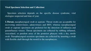
Clinical signs a) Furious form- Anorexia, hiding to isolated dark corner, disobedience to command, involuntary defaecation/urination, sexual excitement, abnormal behaviour like jumping, roaring, bellowing, chewing, biting and dashing at the object, drooling of saliva, paralysis, death b) Dumb form- Yawning, bellowing, roaring, barking, no excitement, inco-ordination, unable to chew or swallow, paralysis, coma ,death PM examination: No gross lesions, non suppurative lesions may be seen Specimen Collection: One half of mid sagittally sectioned brain, cervical spinal cord and parotid salivary gland in 10% formal saline for histopathology Other half of brain in 50% buffered glycerin saline for mice inoculation test, corneal smear and saliva for detection of viral antigen Diagnosis Positive fluorescence antibody test (FAT) GST and Negri bodies in impression smear from brain (Pyramidal or purkinje cells) Mice inoculation test Dot ELISA and Immunohistochemical test Fig.: Negri bodies (Roud/oval, .25 to 27 m
Canine Distemper Etiology It is caused by canine distemper virus (CDV) of genus morbillivirus, family Paramixoviridae Epidemiology Occurs mostly in unvaccinated young carnivores (fox, jackal, wolf, wild dog, hyena, mink, ferret) and also in tigers, snow leopard, jaguars, lions etc Clinical signs Biphasic fever, conjunctivitis, keratitis, photophobia, blindness, hyperkeratosis of foot pads, some times pustules on abdomen Twitching of facial and limb muscles in CNS form Fig. Hard Pad disease
PM findings Thymic atrophy (young pups), necrosis of lymphatic tissues, Bronchopneumonia, enteritis Specimens collections Impression smear from conjunctiva, buffy coat from peripheral blood and formalin fixed specimen of brain, spinal cord, lungs, stomach, intestine, urinary bladder tissues Diagnosis Isolation and identification of virus Demonstration of intranuclear eosinophilic inclusions in respiratory, GI and urinary epithelium Fig. Intranuclear inclusion in Racoon brain
Avian Influenza Etiology Avian influenza or Bird flue is a disease caused by infection with avian influenza type A virus of Orthomyxoviridae Highly pathogenic avian influenza (HPAI) H5N1 in birds N5H8 is highly fatal to wild birds and poultry Epidemiology Duck, geese, pheasants, partridge, psittacine, gulls, shorebirds, emu, eagles and many other species like felids including tiger Pigs play vital role in the interspecies transmission and migratory wild birds specially water fowls are natural host and reservoir Clinical signs Mild and severe respiratory distress, ruffled feathers, depression, decreased feed and water intake in low pathogenic avian influenza (LPAI) HPAI is characterized by respiratory signs, depression, sinusitis, watery diarrhoea, oedema of comb and wattle with cyanosis and haemorrhages, neurological signs and heavy mortality Wild felids show sneezing, nasal discharge, malaise and pyrexia
Differential Diagnosis Botulism, Ranikhet, Pasteurellosis Post Mortem findings Haemorrhages in epicardium, pectoral muscle and proventriculus in poultry Necrotic foci in pancreas, spleen and heart Severe lung congestion with haemorrhages, pleural effusion and serosanguinous exudates in trachea and bronchiolar lumen are found in infected tiger Sample collection Cloacal and oropharyngeal swabs or fresh faeces in virus transport medium Visceral organ stored at -800C or fixed in 10% formalin Diagnosis Detection of virus by egg inoculation, virus isolation method and by RT-PCR BSL- 3 Lab facilities is needed for diagnosis of virus
Miseales Etiology It is caused by measle virus of family paramixoviridae in human and non human primate. Epidemiology Measles is a highly contagious viral disease in nonhuman primates in which infections range from asymptomatic to rapidly fatal, resulting in significant morbidity and mortality in captive populations This has been documented in marmosets, tamarins, owl monkeys etc. and is fatal to them Clinical signs Common marmosets with measles infection become clinically apparent with lethargy, facial edema, and nasal discharge and occasionally develop an exanthema Death occurs 8 18 h after the onset of the first clinical signs. PM findings Necrotizing enterocolitis with hemorrhagic diarrhea
Infectious Canine hepatitis Etiology It is a viral disease caused by canine adenovirus (CAV-1) of adenoviridae family CAV-1 is a relatively tough virus that is able to survive most disinfectants Epidemiology Dog, foxes, wolves and bears are commonly affected Solitary cases of inclusion body hepatitis reported in panther Clinical signs Saddle back fever with leukopenia Blue eye condition (Corneal opacity) Increase blood clotting time and bleeding around deciduous teeth
Diagnosis Signs of abrupt onset and prolonged bleeding Gall bladder with thickened and oedematous wall and necrosis in liver Paint brush haemorrhage in gastric mucosa, lymph nodes, pancreas and sub cutaneous tissues Specimen for lab Formalin fixed liver, kidney, lymph nodes and gastic mucosa Live animals- Whole blood and serum Virus isolation, immuno-flurescence test and presence of characteristics intra- nuclear inclusion in hepatocytes
Kyasanur forest disease (KFD) Kyasanur forest disease (KFD) is a tick-borne viral haemorrhagic fever endemic to South-western part of India Etiology KFD is Caused by Kyasanur Forest disease virus (KFDV), a member of the virus family Flaviviridae Epidemiology Hard ticks (Hemaphysalis spinigera) are the reservoir of KFD virus and once infected, remain so for life Rodents, shrews, and monkeys are common hosts for KFDV after being bitten by an infected tick Monkeys, predominantly, the black-faced langur (Presbytis entellus) and the red faced bonnet monkey (Macaca radiata) are susceptible to KFDV
Clinical signs Incubation period of 3-8 days The symptoms of KFD begin suddenly with chills, fever, and headache Severe muscle pain with vomiting, gastrointestinal symptoms and bleeding problems may occur 3-4 days after initial symptom onset Zoonotic importance Gastrointestinal bleeding complication Diagnosis Molecular detection by PCR or virus isolation from blood Serologic testing using enzyme-linked immunosorbent serologic assay (ELISA) in later stage
Herpes virus infections Etiology Herpes simplex virus type 1 (HSV-1) - oral herpes (affects the mouth and surrounding skin but can also affect the genital region) Herpes simplex virus type 2 ( HSV-2)- typically causes genital herpes, usually sexually transmitted Epidemiology Herpes virus infections are documented in non-human primates, elephant calves etc Rhesus macaques and cynomolgus (crab eating macaque) are considered as the primary natural hosts Clinical signs Lesions in non-human primates are mostly confined to the mucosa of buccal cavity Ulcers or vesicles do occur around the lips and external nares and the most common site is the tongue All macaques are considered to be potential shedders of Cercopithecine (Old world monkey family) herpesvirus type 1 (Herpesvirus simiae B virus) The infection is generally subclinical or mild (conjunctivitis or oral vesicles) in Macaca spp but usually causes a fatal encephalitis and encephalomyelitis in people
Feline Panleukopenia/Feline Distemper It is a highly contagious and life-threatening viral disease in the cat population and characterized by severe dehydration, vomiting, diarrhoea, fever and leukopenia. Etiology Feline parvovirus (FPVis closely related to mink enteritis virus and the type 2 canine parvoviruses (CPV) that cause canine parvoviral enteritis All are now designated as members of the species Carnivore proto-parvovirus 1 Epidemiology FPV can cause disease in all felids and in some members of related families (eg, raccoon, mink), but it does not harm canids Clinical signs Per-acute: Sudden death Acute: Biphasic fever, leukopenia, vomiting, diarrhoea and terminally hyperthermia and death
PM findings No specific lesions, dehydration, dark tarry coloured blood Hyperaemic gastric and intestinal mucosa Necrotic ulcer in ileum and colon Specimen for lab Heparnised whole blood, pieces of intestine, bone marrow and lymph nodes in 10% formalin saline or 50% glycerine saline and on dry ice separately Faecal or rectal swab for virus isolation Diagnosis Isolation of virus and demonstration of characteristic inclusion body in intestinal epithelial cell and histiocytes Kits for Feline panleukopenia antibody detection are available
Parvo virus infection Etiology Several antigenically and genetically very closely related viruses causes Feline pan-leukopenia virus (FPV) Canine parvovirus(CPV-2) along with its antigenic types CPV-2a and CPV-2b Mink enteritis virus(MEV) Blue fox parvovirus (BFPV) Rac-coon parvovirus (RPV) and Raccoon dog parvovirus (RDPV), are grouped informally within the feline parvovirus subgroup
Epidemiology Ingestion of the virus from the environment or by preying on an infected animal Environmental contamination is usually due to viruses that have been shed 4-10 days after infection Parvoviruses are very hardy and can survive for months in cool, moist conditions protected from sunlight and remain viable when frozen Clinical signs Lethargy, depression, and inappetence about four or five days after exposure followed by fever, vomiting, and diarrhea Feces may range from soft to liquid consistency but are typically foul-smelling and may contain mucous or blood
Diagnosis The virus can be detected in feces by ELISA tests or by virus isolation in tissue culture Identification of lesions within the intestines, brain and spinal cord, or lymph tissue during necropsy Canine Parovirus antigen rapid test kit
Feline infectious Peritonitis Feline infectious peritonitis (FIP) is a viral-induced, immune-mediated disease with high fatality rates, affecting domesticated cats and some wild felids globally Etiology Feline infectious peritonitis (FIP) is an immune-mediated disease triggered by infection with a feline coronavirus (FCoV) FCoV belongs to the family Coronaviridae, a group of enveloped, positive-stranded RNA viruses frequently found in cats Epidemiology FCoV infection and FIP occur worldwide with similar prevalence and are found in domestic and wild cats.
Clinical signs Diarrhea and/or vomiting due to replication of FCoV in enterocytes FIP were distinguished 1) An effusive, exudative, wetform : Fibrinous peritonitis, pleuritis, and/or pericarditis with effusion in the abdomen, thorax, and/or pericardium 2) A non-effusive, non-exudative, granulomatous, parenchymatous dry form ; Granulomatous changes in different organs that may include the eyes and CNS 3) A mixed form Diagnosis Peritoneal fluid analysis: The protein content is very high (>3.5 g/dL), consistent with an exudate, whereas the cellular content is low (<5,000 nucleated cells/mL), resembling a modified transudate Cerebrospinal Fluid: Increased protein (50 350 mg/dL with a normal value of <25 mg/dL) and pleocytosis (100 10,000 nucleated cells/mL) PCR has been used to detect FCoV in fecal samples Immunostaining of Feline Coronavirus Antigen
Monkey Pox (Mpox) Mpox is a disease of zoonotic importance Etiology Mpox is caused by mpox virus which is a type of orthopoxvirus of poxviridae. This group of viruses also includes cowpox and smallpox. Epidemiology Evidence of monkeypox virus infection has been found in animals including squirrels, Gambian pouched rats, dormice, different species of monkeys, human and others It can be transmitted through contact with bodily fluids, lesions on the skin or on internal mucosal surfaces Clinical signs Common symptoms of mpox in human are a skin rash or mucosal lesions which can last 2 4 weeks accompanied by fever, headache, muscle aches, back pain, low energy, and swollen lymph nodes Signs and symptoms in animals are not clear and they may act as reservoir Diagnosis Detection of DNA by PCR


")


")


")