Viruses: From Microscopic Intruders to Lethal Agents
Viruses, viroids, and prions - intricate entities challenging our notions of life. Delve into their characteristics, host range, and impact on living organisms to unravel the mysteries of these microscopic intruders.

Download Presentation

Please find below an Image/Link to download the presentation.
The content on the website is provided AS IS for your information and personal use only. It may not be sold, licensed, or shared on other websites without obtaining consent from the author.If you encounter any issues during the download, it is possible that the publisher has removed the file from their server.
You are allowed to download the files provided on this website for personal or commercial use, subject to the condition that they are used lawfully. All files are the property of their respective owners.
The content on the website is provided AS IS for your information and personal use only. It may not be sold, licensed, or shared on other websites without obtaining consent from the author.
E N D
Presentation Transcript
Viruses, Viruses, Viroids Viroids, and Prions , and Prions
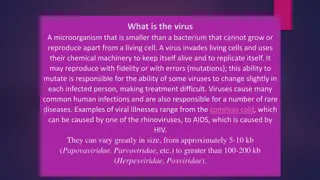
Viruses are too small to be seen with a light microscope and can t be cultured outside their hosts. Advances in molecular biological techniques in the 1980s and 1990s led to the recognition of several new viruses, including human immunodeficiency virus (HIV) and SARS-associated coronavirus.
Are viruses living or non-living organisms? Life can be defined as a complex set of processes resulting from the actions of proteins specified by nucleic acids. The nucleic acids of living cells are in action all the time. Because viruses are inert outside living host cells, in this sense they aren t considered to be living organisms. However, once viruses enter a host cell, the viral nucleic acids become active, and viral multiplication results. In this sense, viruses are alive when they multiply in the host cells they infect. From a clinical point of view, viruses are alive because they cause infection and disease, just as pathogenic bacteria, fungi, and protozoa do.
General Characteristics of Viruses 1. They are small and obligatory intracellular parasites, that is, they absolutely require living host cells in order to multiply. However, both of these properties are shared by certain small bacteria, such as some rickettsias. 2. Viruses contain a single type of nucleic acid (DNA or RNA) and a protein coat, sometimes enclosed by an envelope composed of lipids, proteins, and carbohydrates. 3. No ribosomes 4. They lack enzymes for protein synthesis and ATP generation. To multiply, viruses must take over the metabolic machinery of the host cell.
Host Range Host range refers to the spectrum of host cells in which a virus can multiply. Most viruses infect only specific types of cells in one host species. Host range is determined by the specific attachment site on the host cell s surface and the availability of host cellular factors. Viruses that infect bacteria are called bacteriophages, or phages.
For the virus to infect the host cell, the outer surface of the virus must chemically interact with specific receptor sites on the surface of the cell. For some bacteriophages, the receptor site is part of the cell wall of the host; in other cases, it is part of the fimbriae or flagella. For animal viruses, the receptor sites are on the plasma membranes of the host cells.
Treating diseases with Viruses The potential to use viruses to treat diseases is fascinating because of their narrow host range and their ability to kill their host cells. Experimentally induced viral infections in cancer patients during the 1920s suggested that viruses might have antitumor activity. These tumor-destroying, or oncolytic, viruses may selectively infect and kill tumor cells or cause an immune response against tumor cells. phage therapy: using bacteriophages to treat bacterial infections.
Viral Size Viral Size
Viral Structure Viroid: infectious RNA without capsid that cause some plant diseases, such as potato spindle tuber disease. A virion is an infectious viral particle composed of nucleic acid and surrounded by a protein coat outside a host cell.
Nucleic Acid virus have either DNA or RNA, but never both. The nucleic acid of a virus can be single-stranded or double- stranded. The nucleic acid can be linear or circular. In some viruses (such as the influenza virus), the nucleic acid is in several separate segments.
Viral Nucleic Acid Viral Nucleic Acid
Capsid and Envelope The nucleic acid of a virus is protected by a protein coat called the capsid Each capsid is composed of protein subunits called capsomeres. In some viruses, the capsid is covered by an envelope, which consists of some combination of lipids, proteins, and carbohydrates.
Spikes: Depending on the virus, envelopes may or may not be covered by spikes, which are carbohydrate-protein complexes that project from the surface of the envelope. Some viruses attach to host cells by means of spikes.
Haemagglutination The ability of certain viruses, such as the influenza virus to clump red blood cells is associated with spikes. Such viruses bind to red blood cells and form bridges between them. The resulting clumping is called hemagglutination and is the basis for several useful laboratory tests.
why you can get influenza more than once? why you can get influenza more than once?
General Morphology Viruses may be classified into several different morphological types on the basis of their capsid architecture. 1- Helical viruses resemble long rods that may be rigid or flexible. The viral nucleic acid is found within a hollow, cylindrical capsid that has a helical structure (such as rabies and Ebola) 1- Helical Viruses
2- Polyhedral Viruses Many animal, plant, and bacterial viruses are polyhedral, or many-sided, viruses. The capsid of most polyhedral viruses is in the shape of an icosahedron, a regular polyhedron with 20 triangular faces and 12 corners. Examples of a polyhedral viruses are the adenovirus and the poliovirus.
3- Enveloped Viruses Enveloped viruses are roughly spherical. When helical or polyhedral viruses are enclosed by envelopes, they are called enveloped helical or enveloped polyhedral viruses. Such as the influenza virus and the human herpes virus
4- Complex Viruses Some viruses, particularly bacterial viruses, have complicated structures and are called complex viruses. One example of a complex virus is a bacteriophage.
Taxonomy of Viruses Classification of viruses is based on type of nucleic acid, strategy for replication, and morphology. Virus family names end in -viridae; genus names end in -virus. Family Herpesviridae, genus Simplexvirus. A viral species is a group of viruses sharing the same genetic information and ecological niche. viral species are designated by descriptive common names, such as human immunodeficiency virus (HIV), with subspecies (if any) designated by a number (HIV-1).
Isolation, Cultivation, and Identification of Viruses Viruses must be grown in living cells. The bacteriophages are grown in bacteria. Growing Bacteriophages in the Laboratory The plaque method mixes bacteriophages with host bacteria and nutrient agar. After several viral multiplication cycles, the bacteria in the area surrounding the original virus are destroyed; the area of lysis is called a plaque. Each plaque originates with a single viral particle; the concentration of viruses is expressed as plaque-forming units (PFU).
Growing Animal Viruses in the Laboratory Cultivation of some animal viruses requires whole animals. Some animal viruses can be cultivated in embryonated eggs. In cell cultures virally infected cells are detected via their deterioration, known as cytopathic effects (CPE).
Viral Identification Serological tests: Western blotting (reaction of the virus with antibodies). Nucleic acids: Viruses may be identified by RFLPs and PCR.
Prions Proteinaceous infectious particle, inherited and transmissible by ingestion, transplant, and surgical instruments, first discovered in the 1980s. Prion diseases, such as Creutzfeldt-Jakob disease (CJD) and mad cow disease, all involve the degeneration of brain tissue.
Multiplication of Bacteriophages Bacteriophages can multiply by two alternative mechanisms: the lytic cycle or the lysogenic cycle. The lytic cycle ends with the lysis and death of the host cell, whereas the host cell remains alive in the lysogenic cycle.
Bacteriophage Lambda (): The Lysogenic Cycle Bacteriophage Lambda ( ): The Lysogenic Cycle In contrast to T-even bacteriophages, some viruses don t cause lysis and death of the host cell when they multiply. These lysogenic phages (also called temperate phages) may indeed proceed through a lytic cycle, but they are also capable of incorporating their DNA into the host cell s DNA to begin a lysogenic cycle. In lysogeny, the phage remains latent (inactive). The participating bacterial host cells are known as lysogenic cells.


 Viruses and Bacteria Compared")


: The Lysogenic Cycle")
: The Lysogenic Cycle")