Visual Outcome After Cataract Surgery in Microspherophakia Patients: A Prospective Study
This e-poster presentation discusses the visual outcomes following cataract surgery in patients with microspherophakia, a rare congenital anomaly affecting the crystalline lens. The study examines the surgical procedures and systemic examinations conducted to evaluate the results post-surgery, shedding light on the genetic mutations associated with microspherophakia and the challenges posed by this condition.
Download Presentation

Please find below an Image/Link to download the presentation.
The content on the website is provided AS IS for your information and personal use only. It may not be sold, licensed, or shared on other websites without obtaining consent from the author.If you encounter any issues during the download, it is possible that the publisher has removed the file from their server.
You are allowed to download the files provided on this website for personal or commercial use, subject to the condition that they are used lawfully. All files are the property of their respective owners.
The content on the website is provided AS IS for your information and personal use only. It may not be sold, licensed, or shared on other websites without obtaining consent from the author.
E N D
Presentation Transcript
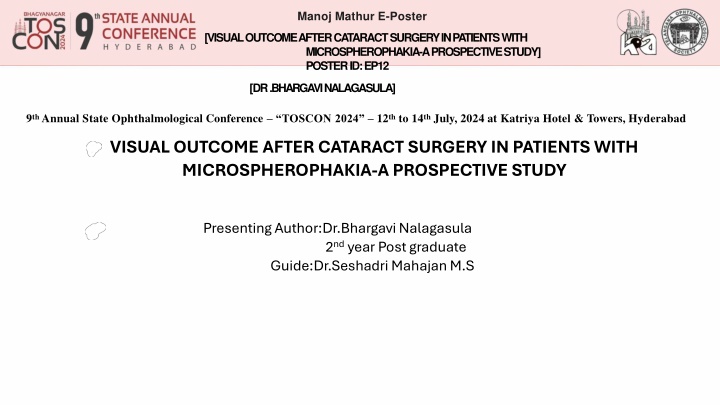
Manoj Mathur E-Poster [VISUAL OUTCOME AFTER CATARACT SURGERY IN PATIENTS WITH MICROSPHEROPHAKIA-A PROSPECTIVE STUDY] POSTER ID: EP12 [DR.BHARGAVI NALAGASULA] 9thAnnual State Ophthalmological Conference TOSCON 2024 12thto 14thJuly, 2024 at Katriya Hotel & Towers, Hyderabad VISUAL OUTCOME AFTER CATARACT SURGERY IN PATIENTS WITH MICROSPHEROPHAKIA-A PROSPECTIVE STUDY Presenting Author:Dr.Bhargavi Nalagasula 2nd year Post graduate Guide:Dr.Seshadri Mahajan M.S
INTRODUCTION: Microspherophakia is a rare congenital anomaly of crystalline lens, bilateral in condition . Characterized by abnormal spherical shape, increased anteroposterior diameter. Homozygous mutation in LTBP2 gene present . Lens dislocation or subluxation, high myopia , Glaucoma may be present. OBJECTIVE: To know visual outcome after cataract surgery in patients with microspherophakia. MATERIALS & METHODS : This is a Prospective study over a period of 5 months from October 2023 to March 2024 at santhiram General Hospital , Nandyal .
OCULAR EXAMINATION: PATIENT-1 PATIENT -2 Right Eye Left Eye Right Eye Left Eye Vision Cf- 3meters Cf-5meters 6/36-6/24 Cf-5 meters 6/60 6/36-6/24 IOP(NCT) 16mmhg 14mmhg 14mmhg 14mmhg Lens NS IV Equator of lens seen NS II Equator of lens seen NS III Central PSCC Equator of lens seen NS II Equator of lens seen Fundus Media hazy Disc seen hazily Media clear 0.4 CDR Vessels -normal Media hazy Disc appears 0.3 CDR Media clear 0.3 CDR Vessels -normal Axial length with in normal limits in both cases A total 2 microspherophakia patients were examined, they underwent Temporal SICS with Iris claw lens under local anaesthesia was done.
SYSTEMIC EXAMINATION: Mandatory to rule out syndromic association RESULTS: POD-1 Patient -1 Patient -2 Vision (RE) UCVA-6/24 with pin hole 6/12 (RE) UCVA-6/12 with pin hole 6/9 IOP(NCT) 14mmHg 12mmHg Adnexa NAD NAD Cornea DM Folds Clear Anterior chamber Quiet Quiet Iris Normal Normal Lens PSPH With Iris claw lens PSPH With iris claw lens Fundus Media clear,0.3 CDR,HNRR, FR+, Vessels -Normal Media clear,0.3 CDR,HNRR, FR+, Vessels -Normal
DISCUSSION: Microspherophakia is a autosomal dominant condition It is due to mutation in latent TGFB binding protein (LTBP2),ADAMTS17,FBN1 genes LTBP2 gene- Protein shows structural homologies with fibrillns , which has a role in regulation of elastic fibre assembly Mutation in LTBP2 gene leads to loss of tension in zonular fibres , making zonular fibres rudimentary , arrest in lens development , remains lens in spherical shape During 5th / 6th of gestational age , nutritional deficiency from defects in Tunica vasculosa causes arrest in development of secondary lens fibres Mutation in ADAMTS 17 gene have shown to disrupt the organization of extracellular matrix, resulting in microspherophakia Microspherophakia associated with systemic conditions like marfans syndrome , will marchesani syndrome , Hyperlysnemia Congenital rubella , Lowe syndrome Patients present with diminution of vision , acute pain , defective accommodation On examination entire equatorial margin of lens is seen with full mydriasis , which will move with change in posture Complications like ectopia lentis , secondary angle closure glaucoma , retinal detachment are seen
CONCLUSION: A complete ophthalmologist, physical examination and genetic advice is recommended for early diagnosis ,timely treatment to avoid complications. REFERENCES: Yu X, Chen W, Xu W. Diagnosis and treatment of microspherophakia. J Cataract Refract Surg. 2020 Dec;46(12):1674-1679. Sauer A, Speeg-Schatz C. [Anterior lens dislocation in the setting of microspherophakia]. J Fr Ophtalmol. 2013 Feb;36(2):186. Shahzadi M, Firasat S, Kaul H, Afshan K, Afzal R, Naz S. Genetic mapping of autosomal recessive microspherophakia to chromosome 14q24.3 in a consanguineous Pakistani family and screening of exon 36 of LTBP2 gene. ]Pak Med Assoc. 2020 Mar;70(3):515-518. Yi H, Zha X, Zhu Y, Lv J, Hu S, Kong Y, Wu G, Yang Y, He Y. A novel nonsense mutation in ADAMTS17 caused autosomal recessive inheritance Weill-Marchesani syndrome from a Chinese family. J Hum Genet. 2019 Jul;64(7):681-687. Simsek T, Beyazyildiz E, Simsek E, zt rk F. Isolated Microspherophakia Presenting with Angle-Closure Glaucoma. Turk J Ophthalmol. 2016Oct;46(5):237-240. [PMC free articlel Muralidhar R, Ankush K, Vijayalakshmi P, George VP. Visual outcome and incidence of glaucoma in patients with microspherophakia. Eye (Lond). 2015Mar;29(3):350-5. [PMC free articlel
