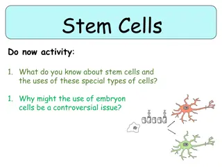
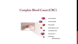
Explore the formation and functions of white blood cells, including eosinophils, basophils, monocytes, and macrophages. Learn about their roles in immunity, response to parasites and allergens, and inflammatory reactions. Discover the intricate processes of maturation from bone marrow to active defense mechanisms in the body.
Please find below an Image/Link to download the presentation.
The content on the website is provided AS IS for your information and personal use only. It may not be sold, licensed, or shared on other websites without obtaining consent from the author. If you encounter any issues during the download, it is possible that the publisher has removed the file from their server.
You are allowed to download the files provided on this website for personal or commercial use, subject to the condition that they are used lawfully. All files are the property of their respective owners.
The content on the website is provided AS IS for your information and personal use only. It may not be sold, licensed, or shared on other websites without obtaining consent from the author.
Lecture content .1 Eosinophils and Basophilophils formation, maturation and function. Monocytes and macrophage formation, maturation and function. Reticuloendothelial system component and function. Lymphocytes formation, maturation and Function. Leucocytosis, leucopenia and leukemia .5 .2 .3 .4 2
Formation and Maturation of Eosinophils Formed in Bone Marrow: 1. Stem cells Myeloblast Promyelocytes 2. Eosinophil myelocytes 3. Eosinophil metamyelocytes 4. polymorphnuclear eosinophil (Mature Eosinophil released to blood) 4
Eosinophil Function Phagocytosis High eosinophil count: Parasitic (hook worm, ascaris, bilharzia) Allergic (asthma, rhinitis, drug reaction) Eosinophil attach themselves to parasites and releases substances (hydrolytic anzymes, superoxide) to kill it. 5
Formation and Maturation of Basophils Formed in Bone Marrow Stem cells Myeloblast Promyelocytes Basophil myelocytes Polymorphnuclear Basophil (Mature Basophils released to blood) .1 .2 .3 7
Basophils Similar to mast. cells secret: Heparin to prevent clotting, Histamine , bradykinin & serotinin contribute to inflammation response The release of those substances cause local and vascular reactions characteristic of allergic manifestation. Mast cell: cell filled with basophil granules, found in connective tissue & releasing histamine & other substances during inflammatory & allergic reactions. 9
Monocytes and Macrophages Formed in Bone Marrow .1 monocytes released into blood. Stay for 10-20 hours in circulation. Then leave blood to tissues transforming into larger cells macrophage. .4 Macrophage life span is long upto few months. Stem cell monoblast promonocyte mature .2 .3 11
Function of Monocytes and Macrophages Macrophages are a powerful phagocytic cells; first line of defense. Ingest up to 100 bacteria, Ingest larger particles as: old RBC Get rid of waste (scanvenger) Functions: anti-inflamatory Directly: phygocytosis of bacteria, dead cells. Indirectly cooperating with lymphocytes by recognizing foreign body (take in foreign body process it and present it to lymphocytes) 12
Reticuloendothelialsystem Consist of: Monocytes. Macrophage. Endothelial cells (bone marrow, spleen, lymph node) Located in all tissues especially: Skin (histocytes), liver (kupffer), spleen, bonemarrow, lymph nodes, lung 16
Functions of Reticuloendothelial system 1. Phagocytosis: Bacterial, dead cells, foreign particles 2. Immune function: processing antigen & antibodies production (indirect) 3. Breakdown of Hb 4. Storage of iron 17
Lymphocytes Formation & Maturation .1 Formed in bone marrow, thymus, lymphoid tissues. Stem cell (thymus, lymphoid tissue & bone marrow) lymphoblast intermediate pyronophilic blast cell lymphocytes .3 Life Span Of Lymphocytes ranges from weeks to months according to the type. .2 19
T-Lymphocytes (Thymus dependent) Formed in bone marrow or lymphoid tissue migrate to thymus for maturation. Life spans 100-300 days. Circulate between blood, tissues, lymph. Types of T-lymphocytes T-helper T-cytotoxic Natural killer Functions Cellular immunity (graft rejection - delayed hypersensitivity.) Role in antibody secretion. 21
B- Lymphocytes (thymus-independents) First discovered in Bird Bursa Formed in: Bone marrow, germinal layer of lymph node, red pulp of spleen Life span 2-7 days Stimulated by antigen It transforms into largeplasma cell (produce antibody) Function: Humoral immunity. 23
Types of Immunity Immune system Innate (non-specific; natural) immunity Adaptive (specific; acquired) immunity 1. Third line of defense. 2. Antigen specificity. It is activated by thousands of diverse antigens. 3. Responds with the proliferation of cells and the generation of antibodies. 4. Responds slowly, being fully activated about 4 days after the immunologic threat. 5. Exhibits immunologic memory, so that repeated exposure to the same infectious agent results in improved resistance against it. 1. Second line of defense 2. Is present at birth 3. Persists throughout life 4. Can be mobilized rapidly and act quickly 5. Attacks all antigens fairly equally
The Complement System is Part of the 1stLine of Defense complement system: 1- The first part of the immune system that meets invaders such as bacteria 2-it is a group of proteins. 3- These proteins flow freely in the blood and can quickly reach the site of an invasion where they can react directly with antigens ( molecules that the body recognizes as foreign substances). functions of complement proteins (When activated ): 1. Trigger inflammation. 2. Attract eater cells such as macrophages to the area. 3. Coat intruders so that eater cells are more likely to devour (swallow and eat) them (a process called as opsonization). 4. Kill intruders.
Leucocytosis Increased number of WBC Physiological Diurnal morning evening After physical exercise Stress or Adrenaline injection Pathological: Bacterial infection (tonsillitis, Appendicitis) Worm infection. Allergric reactions. 30
Leucopenia Deficiency of the white blood cells: Causes: 1- malnutrition. 2- typhoid fever. 3- drugs. 4- B12 & folic acid 5- radiation 31
Leukaemia Cancer of white cells due to chromosomal abnormality caused by chemicals, radiation, and viruses. WBC more than 50x103 Types of leukaemia Myeloblast leukaemia myeloid cells Lymphoblast leukaemia lymphocytic cells Acute or chronic onset Accompanied with anaemia, bleeding 32
Objectives At the end of this lecture student should be able to: Describe Esinophils formation and .1 functions Describe Basophils formation and .2 functions .3 Describe Monocytes and macrophage formation and functions. Describe Reticuloendothelial componants .4 and functions 33
Objectives At the end of this lecture student should be able to: 5. Describe lymphocytes formation and maturation. 6.Describe the functions of the different types of lymphocytes. 7. Recognise leucocytosis and leucopenia. 8. Recognize type of leukaemia 34