X-ray Production and Imaging
X-ray production involves accelerating electrons from a cathode to anode in an X-ray tube. The process results in the emission of X-ray radiation, which is used in conventional X-ray imaging to visualize internal structures. By increasing the kinetic energy of electrons through high electrical potential, the intensity and quality of the X-ray beam are enhanced. Learn about the interaction between electrons and targets in X-ray tubes for efficient production of diagnostic images.
Download Presentation

Please find below an Image/Link to download the presentation.
The content on the website is provided AS IS for your information and personal use only. It may not be sold, licensed, or shared on other websites without obtaining consent from the author.If you encounter any issues during the download, it is possible that the publisher has removed the file from their server.
You are allowed to download the files provided on this website for personal or commercial use, subject to the condition that they are used lawfully. All files are the property of their respective owners.
The content on the website is provided AS IS for your information and personal use only. It may not be sold, licensed, or shared on other websites without obtaining consent from the author.
E N D
Presentation Transcript
X-ray Production (Chapter-4) BY BY DR.SALIH DR.SALIH OMER OMER ( (202 2022 2- -2023 2023) ) 4 4th th- - year Medical year Medical
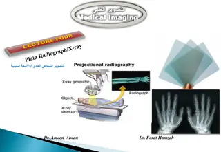
Conventional X-ray Imaging. X-ray Production. High Electrical Potential Electrons + - Electrons from cathode filament are accelerated towards and impact the rotating anode. Rapid deceleration produces heat ( ~ 98 %) and x-rays (~ 2%) Radiation Penetrate the Sample Exposure Recording Device
Overcouch X-ray Tube and Table High Tension Cables X-ray Tube housing Controls Light Beam Diaphragm Table, and cassette holder
How are X-Rays Produced? X-Rays are produced in a special type of tube called An X ray Tube!!
Electrons are first emitted from a heated filament, by a process called thermionic emission. They are then accelerated across the evacuated X-ray tube, under the action of a large voltage across the tube, the filament forming the negative cathode and the target being positive anode. On striking the target, the electrons lose most (about 99%) of their energy in low-energy collisions with target atoms, resulting in a substantial heating of the target. The rest of the electron energy (usually less than 1%) reappears as X-ray radiation.
A rapidly-rotating anode is generally used. It forms the (tungsten) target surface on to which the electron beam is focused. The target area under bombardment is constantly changing, thus reducing local heat concentration. (You can often hear the whirring of the anode motor during the taking of an X-ray. Copper, being an excellent heat conductor, is used to hold the anode in place. Oil, which circulates in the outer housing,, helps with convective cooling (as well as providing electrical insulation).
ELECTRON TARGET INTERACTIONS: The x-ray imaging system description in Chapter 3 emphasizes that its primary function is to accelerate electrons from the cathode to the anode in the x-ray tube. The three principal parts of an x-ray imaging system the operating console, the high-voltage generator, and the x-ray tube are designed to provide a large number of electrons with high kinetic energy focused toward a small spot on the anode. Kinetic Energy is the energy of motion. In determining the magnitude of the kinetic energy of a projectile, velocity is more important than mass. In an x-ray tube, the projectile is the electron. All electrons have the same mass; therefore, electron kinetic energy is increased by raising the peak Kilovoltage (kVp). As electron kinetic energy is increased, both the intensity (quantity) and the energy (quality) of the x-ray beam are increased.
ELECTRON TARGET INTERACTIONS: Electrons traveling from cathode to anode constitute the x-ray tube current and are sometimes called projectile electrons. When these projectile electrons hit the heavy metal atoms of the x-ray tube target, they transfer their kinetic energy to the target atoms. These interactions occur within a very small depth of penetration into the target. As they occur, the projectile electrons slow down and finally come nearly to rest, at which time they are conducted through the x-ray anode assembly and out into the associated electronic circuitry. The projectile electron interacts with the orbital electrons or the nuclear field of target atoms. These interactions result in the conversion of electron kinetic energy into thermal energy (heat) and electromagnetic energy in the form of infrared radiation (also heat) and x-rays.
Figure.1 Most of the kinetic energy of projectile electrons is converted to heat by interactions with outer-shell electrons of target atoms. These interactions are primarily excitations rather than ionizations.
Anode Heat: Approximately 99% of the kinetic energy of projectile electrons in converted to heat. So only 1% of the kinetic energy of electrons is used for the production of x- radiation. Therefore, sophisticated as it is, the x-ray imaging system is very inefficient. The production of heat in the anode increases directly with increasing x-ray tube current. Doubling the x-ray tube current doubles the heat produced. Heat production also increases directly with increasing kVp, at least in the diagnostic range. Although the relationship between varying kVp and varying heat production is approximate, it is sufficiently exact to allow the computation of heat units for use with anode cooling charts. The efficiency of x-ray production is independent of the tube current. Consequently, regardless of what mA is selected, the efficiency of x-ray production remains constant. The efficiency of x-ray production increases with increasing kVp. At 60 kVp, only 0.5% of the electron kinetic energy is converted to x-rays. At 100 kVp, approximately 1% is converted to x-rays, and at 20 MV, 70% is converted.
Characteristic Radiation If the projectile electron interacts with an inner-shell electron of the target atom rather than with an outer-shell electron, characteristic x-rays can be produced. Characteristic x-rays result when the interaction is sufficiently violent to ionize the target atom through total removal of an inner-shell electron. The transition of an orbital electron from an outer shell to an inner shell is accompanied by the emission of an x-ray. The x-ray has energy equal to the difference in the binding energies of the orbital electrons involved.
Bremsstrahlung Radiation: The production of heat and characteristic x-rays involves interactions between the projectile electrons and the electrons of x-ray tube target atoms. A third type of interaction in which the projectile electron can lose its kinetic energy is an interaction with the nuclear field of a target atom. In this type of interaction, the kinetic energy of the projectile electron is also converted into electromagnetic energy. Because the electron is negatively charged and the nucleus is positively charged, there is an electrostatic force of attraction between them As the projectile electron passes by the nucleus, it is slowed down and changes its course, leaving with reduced kinetic energy in a different direction. This loss of kinetic energy reappears as an x-ray.
These types of x-rays are called bremsstrahlung x-rays. Bremsstrahlung is a German word that means slowed-down radiation. Bremsstrahlung x-rays can be considered radiation that results from the braking of projectile electrons by the nucleus. Figure 2. Bremsstrahlung x-rays result from the interaction between a projectile electron and a target nucleus. The electron is slowed, and its direction is changed.
X-RAY EMISSION SPECTRUM X-ray emission spectra have been measured for all types of x-ray imaging systems. Data on x-ray emission spectra are needed if one is to gain an understanding of how changes in kVp, mA, and added filtration affect the quality of an image. Figure. 3 General form of an x-ray emission spectrum.
Factors that influence the shape of X-ray Emission spectra 1- the projectile electrons accelerated from the cathode to the anode not all have the peak kinetic energy. Depending on the type of rectification and high-voltage generation, many of these electrons may have very low energies when they strike the target. Such electrons can produce only low-energy x-rays. 2- The target of a diagnostic x-ray tube is relatively thick. Consequently, many of the bremsstrahlung x-ray emitted result from multiple interactions of the projectile electrons, and for each successive interaction, a projectile electron has less energy. 3- Low-energy x-rays are more likely to be absorbed in the target. 4- External filtration is nearly always added to the x-ray tube assembly. This added filtration selectively removes low-energy x-ray from the beam.
Factors Affecting The X-Ray Emission Spectrum 1-Effect of mA and mAs: If one changes the current from 200 to 400 mA while all other conditions remain constant, twice as many electrons will flow from the cathode to the anode, and the mAs will be doubled. This operating change will produce twice as many x-rays at every energy. In other words, the x-ray emission spectrum will be changed in amplitude but not in shape Note: A change in mA or mAs results in a proportional change in the amplitude of the x-ray emission spectrum at all energies.
Factors Affecting The X-Ray Emission Spectrum 1-Effect of KVp: As the kVp is raised, the area under the curve increases to an area approximating the square of the factor by which kVp was increased. Accordingly, the x-ray quantity increases with the square of this factor. When kVp is increased, the relative distribution of emitted x-ray energy shifts to the right to a higher average x-ray energy. The maximum energy of x-ray emission always remains numerically equal to the kVp Note: Change in kVp results in an increase in the amplitude of the emission spectrum at all energies but a greater increase at high energies than at low energies. Therefore, the spectrum is shifted to the right, or high-energy, side.
Figure 4. demonstrates the effect of increasing the kVp while other factors remain constant. The lower spectrum represents x-ray operation at 72 kVp, and the upper spectrum represents operation at 82 kVp a 10-kVp (or 15%) increase
A 15% increase in kVp does not double the x-ray intensity but is equivalent to doubling the mAs to the image receptor. To double the output intensity by increasing kVp, one would have to raise the kVp by as much as 40%. Radiographically, only a 15% increase in kVp is necessary because with increased kVp, the penetrability of the x-ray beam is increased. Therefore, less radiation is absorbed by the patient, leaving a proportionately greater number of x-rays to expose the image receptor.
Factors Affecting The X-Ray Emission Spectrum 3-Effect of Added Filtration: Adding filtration to the useful x-ray beam reduces x-ray beam intensity while increasing the average energy. This effect is shown in Figure 5., where an x-ray tube is operated at 95 kVp with 2-mm aluminum (Al) added filtration compared with the same operation with 4-mm Al added filtration. Added filtration more effectively absorbs low-energy x-rays than high-energy x-rays; therefore, the bremsstrahlung x-ray emission spectrum is reduced further on the left than on the right. Adding filtration is sometimes called hardening the x-ray beam because of the relative increase in average energy. The characteristic spectrum is not affected, nor is the maximum energy of x-ray emission. There is no simple method for calculating the precise changes that occur in x-ray quality and quantity with a change in added filtration.
Figure 5. Adding filtration to an x-ray tube results in reduced x-ray intensity but increased effective energy. The emission spectra represented here resulted from operation at the same mA and kVp but with different filtration.
Factors Affecting The X-Ray Emission Spectrum 4-Effect of Target Material: The atomic number of the target affects both the number (quantity) and the effective energy (quality) of x-rays. As the atomic number of the target material increases, the efficiency of the production of bremsstrahlung radiation increases, and high-energy x-rays increase in number to a greater extent than low-energy x-rays. Note: Increasing target atomic number enhances the efficiency of x-ray production and the energy of characteristic and bremsstrahlung x-rays.
These changes are shown schematically in Figure 6. Tungsten is the primary component of x-ray tube targets, but some specialty x-ray tubes use gold as target material. The atomic numbers for tungsten and gold are 74 and 79, respectively. Molybdenum (Z = 42) and rhodium (Z = 45) are target elements used for mammography. In many dedicated mammography imaging systems, these elements are incorporated separately into the target. The x-ray quantity from such targets is low owing to the inefficiency of x-ray production. This occurs because of the low atomic number of these target elements. Elements of low atomic number also produce low-energy characteristic x-rays. Figure 6. Discrete emission spectrum shifts to the right with an increase in the atomic number of the target material
Factors Affecting The X-Ray Emission Spectrum 5-Effect of Voltage wave form: There are five voltage waveforms: half-wave rectified, full-wave rectified, three-phase/six-pulse, three-phase/ 12-pulse, and high-frequency waveforms. Half-wave rectified and full-wave rectified voltage waveforms are the same except for the frequency of x-ray pulse repetition. There are twice as many x-ray pulses per cycle with full-wave rectification as with halfwave rectification. The difference between three- phase/six-pulse and three-phase/12-pulse power is simply the reduced ripple obtained with 12-pulse generation compared with six-pulse generation. High- frequency generators are based on fundamentally different electrical engineering principles. They produce the lowest voltage ripple of all high-voltage generators
Figure.7 Three-phase and high-frequency operations are considerably more efficient than single-phase operation. Both the x-ray intensity (area under the curve) and the effective energy (relative shift to the right) are increased. Shown are representative spectra for 92- kVp operation at constant mAs. The x-ray emission spectrum that results from high-frequency operation is more efficient than that produced with a single-phase or a three-phase generator. The area under the curve is considerably greater, and the x-ray emission spectrum is shifted to the high-energy side. The characteristic x-ray emission spectrum remains fixed in its position on the energy axis but increases slightly in magnitude as a result of the increased number of projectile electrons available for K-shell electron interactions.
Q1. An interventional radiology system is capable of 1200mA when operated in 100kVp, 100ms. What is the power rating? Q2. A single-phase radiographic unit installed in a private office reaches maximum capacity at 100 ms of 120 kVp and 500 mA. What is its power rating? Q3. Suppose the curve labeled 72 KVp covers a total area of 3.6cm2 and represents an x-ray quantity of 125 mR(1.25 mGy). What area under the curve and x-ray quantity would be expected for operations at 82 KVp?
SUMMARY When electrons are accelerated from the cathode to the target of the anode, three effects take place: the production of heat, the formation of characteristic x-rays, and the formation of bremsstrahlung x-rays. Characteristic x-rays are produced when an electron ionizes an inner-shell electron of a target atom. As the inner-shell void is filled, a characteristic x-ray is emitted. Bremsstrahlung x-rays are produced by the slowing down of an electron by the target atom s nuclear field. Most x-rays in the diagnostic range (20 150 kVp) are bremsstrahlung x-rays. X-ray emission spectra can be graphed as the number of x-rays for each increment of energy in keV. Characteristic x-rays of tungsten have a discrete energy of 69 keV. Bremsstrahlung x-rays have a range of energies up to X keV, where X is the kVp. The following four factors influence the x-ray emission spectrum: (1) low-energy electrons interact to produce low-energy x-rays, (2) successive interactions of electrons result in the production of x-rays with lower energy, (3) low-energy x-rays are most likely to be absorbed by the target material, and (4) added filtration preferentially removes low-energy x-rays from the useful beam.


