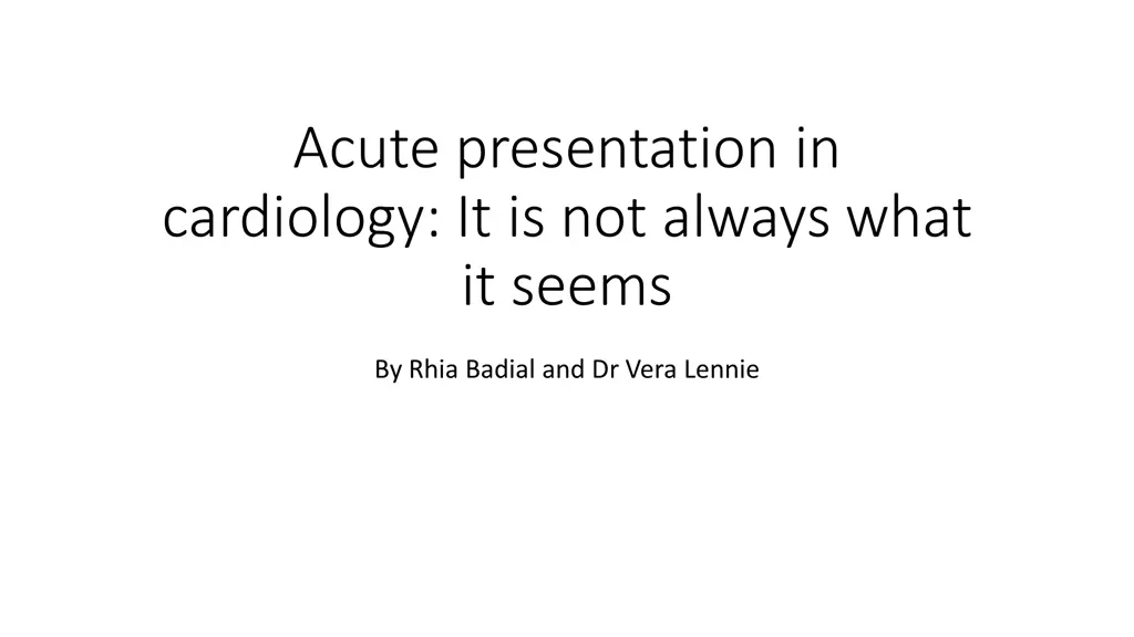
Acute presentation in cardiology: It is not always what it seems
Gain insights into different causes of chest pain and how to construct a differential diagnosis using history and examination. Understand the significance of elevated troponin and the impact of COVID-19 in cardiology.
- Acute presentation
- cardiology
- chest pain
- differential diagnosis
- elevated troponin
- investigations
- COVID-19
- causes

Download Presentation

Please find below an Image/Link to download the presentation.
The content on the website is provided AS IS for your information and personal use only. It may not be sold, licensed, or shared on other websites without obtaining consent from the author. Download presentation by click this link. If you encounter any issues during the download, it is possible that the publisher has removed the file from their server.
E N D
Presentation Transcript
Acute presentation in cardiology: It is not always what it seems By Rhia Badial and Dr Vera Lennie
Learning outcomes Develop understanding of different causes of chest pain Use history and examination to construct a differential diagnosis Understanding differential diagnoses of elevated troponin Identify relevant investigations to aid diagnosis Consider the impact of Covid19 in cardiology
Presenting complaint 35 year old black man referred from the GP to cardiology 2 episodes of retrosternal chest pain He says he is struggling to catch his breath Recently he has also been experiencing dizzy spells, headaches and tiredness General weakness over the last few days
History of presenting complaint Chest pain described as a tightness No radiation Pain lasts for around 1 minute Both episodes of chest pain occurred on rest Ibuprofen improves the pain slightly He has not experienced any palpitations, ankle swelling or claudication
No previous hospital admissions No cardiac family history Non-smoker, non-drinker and no illicit drug use On no medications Normally very well and sporty
Due to Covid19 what other information may be relevant?
Has none of the main Covid19 symptoms or any other infective symptoms (fever, cough or loss of taste and smell) and has tested negative No family members unwell Has not had either of the Covid19 vaccines Travels to London regularly for work
Examination Normal heart sounds Reduced air entry in left upper zone of chest Warm and well perfused peripheries Capillary refill time < 2 seconds No peripheral oedema Calves soft and non tender No lymphadenopathy Apyrexial
Initial Investigations? ECG EEG Chest xray Blood tests ECHO CT scan Cardiac angiogram
Results ECG T wave inversion in V2 to V6 and biphasic ST-T segment elevation in V3 Elevated troponin 2021 CRP < 1 Normal white cell count Normal renal and liver function Normal chest xray ECHO no left ventricular damage, no hypertrophy and no valvular abnormalities
Cardiac Angiogram Normal coronary arteries with no evidence of stenosis or occlusion, which could result in coronary spasm or myocardial infarction
What can cause elevated troponin? Myocardial infarction Arrythmia Pulmonary embolism Septic shock Aortic dissection Cholecystitis Cocaine Myocarditis Percutaneous coronary intervention (PCI)
Causes of elevated troponin Any infection resulting in inflammation can elevate troponin levels, the troponin levels can also elevate and reduce daily due to random cytokine responses.
Cardiac MRI Cardiac MRI revealed inflammation around the heart muscle and patient was diagnosed with myocarditis Most likely combined with pericarditis Sometimes this is referred as myopericarditis
What are the typical ECG features in pericarditis? Widespread concave ST elevation Delta wave PR depression Hyperacute T wave Dynamic ST changes Sinus tachycardia
Patients ECG 4 weeks after admission Concave ST elevation
Diagnostic criteria for myocarditis History Examination Initial investigations: ECG, troponin and chest xray Coronary angiogram Cardiac MRI Consider endomyocardial biopsy in required
Further investigations and management Enforced rest Statins Dalteparin Give 500mls Plasmalyte 148 ACE inhibitor Administer 15l Oxygen Analgesia Viral screen Serum ACE
Monitoring Continuous ECG monitoring during acute phase Daily troponins It is important for the patient to undergo regular ECHOs to ensure their left ventricular function is persevered it should be > 55% however ideally should be compared to previous ones to see if it has reduced from the patient s normal Cannot discharge patient until their troponin has reduced to normal due to risk of arrhythmias
Long term complications of myocarditis Fibrosis leading to left ventricular impairment Ventricular arrythmias Chronic hypoxia Life threatening Angina on exertion Heart failure
Complications of long Covid19 Pneumonia Acute respiratory distress syndrome Bacterial infection Primary cancer Myocarditis Infertility Septic shock Multi organ failure Permanent paralysis Cognitive impairment Obesity Breathlessness
Summary Myocarditis can present with non-specific symptoms There can be no obvious cause for myocarditis it was believed that this patient developed it due to a previous episode of undiagnosed Covid19 (which is now recognised as a common cause) Troponin can be elevated by many factors other than myocardial infarction ECG can have evidence of ST elevation in myocarditis The treatment for myocarditis is mainly supportive Covid19 can cause a range of long-term complications
Further Reading You have now: Developed your understanding of non acute coronary causes of chest pain Used history and examination to construct a differential diagnosis Learnt to identify abnormalities on ECGs Reviewed management of myocarditis Further Reading More detailed learning on myocarditis: European Society of Cardiology Link between myocarditis and Covid19 vaccine: BMJ