Chest Imaging Interpretation for Tuberculosis Screening

Chest imaging plays a crucial role in screening for pulmonary tuberculosis (TB). This activity, led by Dana Kissner from Wayne State University Detroit Tuberculosis Clinic, focuses on interpreting chest imaging results to aid in the diagnosis and management of TB cases. The content includes information on radiological terms used in TB cases, the importance of chest radiography, and case studies illustrating TB screening and treatment strategies. Participants can earn continuing education credits through this informative session.

Chest Imaging Interpretation for Tuberculosis Screening
PowerPoint presentation about 'Chest Imaging Interpretation for Tuberculosis Screening'. This presentation describes the topic on Chest imaging plays a crucial role in screening for pulmonary tuberculosis (TB). This activity, led by Dana Kissner from Wayne State University Detroit Tuberculosis Clinic, focuses on interpreting chest imaging results to aid in the diagnosis and management of TB cases. The content includes information on radiological terms used in TB cases, the importance of chest radiography, and case studies illustrating TB screening and treatment strategies. Participants can earn continuing education credits through this informative session.. Download this presentation absolutely free.
Presentation Transcript
Lab test Interpretation: Chest imaging Dana Kissner Wayne State University Detroit Tuberculosis Clinic
2023 MDHHS World TB Day March 24, 2023 Disclosure No one involved in the planning or presentation of this activity has any relevant financial relationships to disclose. WesternMichigan University Homer Stryker M.D. School of Medicine adheres to the ACCME s Standards for Integrity and Independence in Accredited Continuing Education. Any individuals in a position to control the content of a CE activity, including faculty, planners, reviewers or others are required to disclose all relevant financial relationships with ineligible entities (commercial interests). All relevant conflicts of interest have been mitigated prior to the commencement of the activity. Accreditation In support of improving patient care, this activity has been planned and implemented by WesternMichigan University Homer Stryker M.D. School of Medicine and MDHHS. Western Michigan University Homer Stryker M.D. School of Medicine is jointly accredited by the Accreditation Council for Continuing Medical Education (ACCME), the Accreditation Council for Pharmacy Education (ACPE), and the American Nurses Credentialing Center (ANCC), to provide continuing education for the healthcare team. Credit amount subject to change. Interprofessional Continuing Education This activity was planned by and for the healthcare team, and learners will receive 5.0 Interprofessional Continuing Education (IPCE) credits for learning and change. Physicians WesternMichigan University Homer Stryker M.D. School of Medicine designates this live activity for a maximum of 5.0 AMA PRA Category 1 Credits . Physicians should claim only the credit commensurate with the extent of their participation in the activity. Nurses WesternMichigan University Homer Stryker M.D. School of Medicine designates this activity for 5.0 contact hours for nurses. Nurses should claim only credit commensurate with the extent of their participation in the activity.
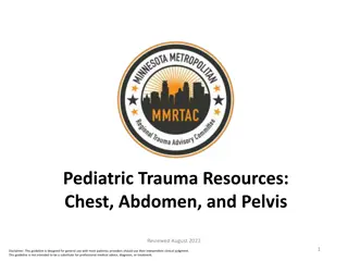
Transmission 10% Airborne droplets Progressive primary tuberculosis Primary tuberculosis 90% CXR may be abnormal due to something something else. else. 5 - 10% Active tuberculosis (disease) Latent tuberculosis infection
Chest radiography, or chest X-ray (CXR), is an important tool for triaging and screening for pulmonary TB, and it is also useful to aid diagnosis when pulmonary TB cannot be confirmed bacteriologically. 2016 2016
Terms used by radiologists in 97 chest reports: 38 Consecutive TB cases 2017- March 24, 2021 24/97 24/97
Case 1: (Like Dr. Dicksons case 1) An 18-year-old man from China is a new student at a college in the Detroit region. PPD (TB) skin test was positive. IGRA not done due to cost. After treatment completion, IGRA was done and was negative. He answers no to having symptoms. Further questioning reveals: 1. He has always been under weight. 2. He coughs, but attributes it to allergies.
Red arrows -nodule, opacity, density Blue arrow - linear, nodular, fibrotic, old, healed, scar Yellow arrows calcification, can be ignored Note upper lobe location Primary TB usually mid to lower lobe disease Upper lobe, superior segment of lower lobe are common sites for reactivation TB. TB can be anywhere in lung
Look at CXR in context of TB risk factors (China) and carefully obtained medical history (underweight, cough) Negative IGRA does not exclude TB! Patient has symptoms of TB & needs CXR and sputum tests Screening for risk factors & symptoms is key Next step is to collect adequate sputum. Treat for TB after adequate sputum is collected. And complete treatment.
Percentage TB Cases by Case Verification Criteria, United States, 2021 (N=7,882) Positive culture 79% Positive IGRA or PPD + clinical/radiographic findings No microbiologic confirmation Clinical case 13% Clinical &/or radiographic findings only & treated for TB disease No microbiologic confirmation Provider diagnosis 4% Positive NAA* test 3% Positive smear <1% *NAA=nucleic acid amplification
Case 2: Dr. Dicksons case annotated A 47-year-old male with a history of living in shelters has hemoptysis and unexplained weight loss. Screening has already been done by symptom review Should he be screened? No. Done Should he be tested? Yes. + Chest x-ray + Sputum +/- IGRA? Yes, but least essential
Cavities caused by TB: Air filled oval or round spaces Walls are thin to moderate in thickness Contain little to no fluid Contain many rapidly reproducing TB organisms Often highly contagious
Cavities: Other TB examples Cavity forming in consolidation Consolidation: homogeneous translucent opacity, made up of fluid, cells, pus, or blood Overlapping cavities Air-filled bronchi form air bronchograms
Cavities: Other examples Cavity forming in consolidation Consolidation: homogeneous opacity, made up of fluid, cells, pus, or blood Circles are cavities Arrows are air bronchograms in consolidation Overlapping cavities Air-filled bronchi form air bronchograms
Case 3: A 35 year old woman, in septic shock in ICU Fever of 40 degrees C Low blood pressure High heart rate Elevated lactic acid While in prison 10 years ago, she had a positive PPD, untreated
Miliary TB Small nodule is old, probably the initial focus of her TB (red arrow) Multiple small nodules, uniform in size 1-5 mm Miliary TB is difficult to see early on A medical emergency Resembles millet seeds
Case 4: A 55-year-old alcoholic is short of breath. He has had weight loss and cough over the past month. Arrows show free flowing fluid forming a meniscus
Pleural effusion Can be 1st sign of primary TB More opaque than consolidation Fluid is exudative (high protein, LDH) in TB Effusion can be small to massive, yellow to bloody, free flowing or loculated Fluid should be tapped (thoracentesis) and tested for TB
Case 5: 15-year-old boy from Africa with difficulty swallowing, weight loss, fevers, and sweats. Yellow arrow narrowed esophagus Purple arrow narrowed right mainstem bronchus
Mediastinal & hilar lymphadenopathy Frequently seen in primary TB Can surround and close off structures such as esophagus Can erode into heart, esophagus, trachea, and bronchi Blue arrow points to dye lighting up (bright white) rim of the lymph nodes, surrounding air in the middle caused by necrosis (cell death). Radiologists use the term rim enhancement and low attenuation centrally.
Case 5: A 60 year man complains of cough, sputum production, loss of appetite, weight loss, night sweats, and hemoptysis Tree-in-bud pattern Seen best with high resolution CT scans


, is")

 An 18-year-old man")
 and")