Acute Respiratory Failure involves various physiological mechanisms resulting in compromised lung functions leading to hypoxemia and hypercapnia. Learn about the main physiological values, non-respiratory functions of the lung, classification of ARF, types of hypoxia, and the management of acute pulmonary failure. Explore causes of ventilatory failure and different types of hypoxia in this comprehensive guide.
Please find below an Image/Link to download the presentation.
The content on the website is provided AS IS for your information and personal use only. It may not be sold, licensed, or shared on other websites without obtaining consent from the author. If you encounter any issues during the download, it is possible that the publisher has removed the file from their server.
You are allowed to download the files provided on this website for personal or commercial use, subject to the condition that they are used lawfully. All files are the property of their respective owners.
The content on the website is provided AS IS for your information and personal use only. It may not be sold, licensed, or shared on other websites without obtaining consent from the author.
Physiology of the Respiration Non- respiratory functions of the lung 1. Blood filter 2. Participation to electolite & water balance 3. Production of protein & fat molecules (surfactant) 4. Participation in thermogenesis & thermolysis 5. Hemodynamic balance between right and left heart 6. Component of ABB 7. Control under various mediators (hormones, kinines, etc.) 8. Part of hemostasis and fibrinolitic systems 9. Part of the immune system
Physiology of the Respiration Main physiologic mechanisms involved in Acute Respiratory Failure 1. Restrictive (decrease in the number of functional lung units) 2. Obstructive (disturbances of air passage to alveoli) 3. Impaired diffusion through ACM 4. Impaired perfusion 5. Ventilation-Perfusion mismatch 6. Alveolar shunting 7. Dead Space
Physiology of the Respiration Some physiological values (normal range) Rate Resp.min.volume(cm3/kg min) Tidal volume (cm3/kg) Vital capacity (cm3/kg) PaO2 mm Hg PvO2 mm Hg PaCO2 mm Hg Vd/Vt Qs/Qt % 12-16 /min 70-130 6-8 60-70 90-100 37- 42 36 44 0.3 0.35 less than 7
Respiratory Failure Five types of hypoxia 1. Respiratory (pulmonary) 2. Circulatory (impaired micro-circulation) 3. Hematic (impaired oxygen binding capacity of hemoglobin) 4. Tissue (inability of the cell enzymes to use oxygen cyanic and CO) 5. Combined
Management of Acute Pulmonary Failure I. Non-specific measures A. Free Airway B. Optimization of the inhaled gas blend C. Artificial Ventilation II. Specific measures
Ventilatory Failure Most common causes: A). Pharmacological 1. Muscle relaxants ( diaphragmatic paralysis) 2. Narcotics (direct respiratory depression) B). Neuromuscular Diseases C). Limitations to Diaphragmatic Excursion 1. Postoperative 2. Obesity 3. Intestinal obstruction 4. Supine position 5. Bandages or casts D). Flail Chest (ribs fractured in two places) E). Hydro/Hemo/Pneumo Thorax F). Asthma
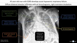
Respiratory Failure The most common causes: 1. Pulmonary Edema 2. Atelectasis 3. Infection (pneumonia, especially massive) 4. Embolism 5. Aspiration 6. Pulmonary Contusion 7. Massive Hemoptysis 8. Pulmonary Burns 9. Interstitial Fibrosis
Management of Status Asthmaticus 1. Check air movement, cardiac status & oxygenation 2. R/o pulmonary embolus, pulmonary edema, pneumothorax, or mucous plugs 3. Nasal O2 early 4. Albuterol (or equivalent) 5. Epinephrine 0.3 ml (1:1000) SQ 6. Theophylline (500 mg/500 ml D5W or D5 NS); Loading dose 6 mg/kg, then 0.9 mg/kg/min 7. Methylprednisolone 40-60 mg q6h/ Solucortef 100 200 mg q4h 8. Monitor ABG s (watch for increase in pCO2) 9. D5 NS with potassium supplementation for hydration 10. Pulmonary toilet
PULMONARY EDEMA is increased extravascular lung water due to a rate of fluid and protein transport from the intravascular space that exeeds the normal clearance mechanisms of the interstitium
PULMONARY EDEMA Symptoms: dyspnea orthopnea paroxysmal noctural dyspnea fatigue nausea cough agitation loss of consciousness
PULMONARY EDEMA Signs: rales tachypnea, cyanosis tachycardia Chest X ray reveal interstitial or alveolar pulmonary edema ABG s hypoxemia usually with a normal or low pCO2
PULMONARY EDEMA Management of Acute Pulmonary Edema 1. Assess vital signs rapidly: pulse, respiratory rate and BP. Perform a rapid physical examination looking for: cyanosis, status of pripheral perfusion, jugular venous distension, abnormal heart and lung sounds & peripheral pulses 2. Administer O2 either by mask or by endotracheal intubation 3. Obtain ABG s 4. Elevate head of the bed 5. Establish a reliable IV line and place foley catheter 6. Obtain: ECG, CXR, electrolites, blood sugar, BUN, creatinine, type blood and complete blood count 7. Establish continuous ECG monitoring, CVP, PCWP
Management of Acute Pulmonary Edema 8. Begin non-specific treatment: - Morphine Sulfate - Furosemide - Nitroglycerine/Nitroprusside 9. Specific underlying precipitating factors such as arrhythmias, fluid overload, extent of pre-existing CHF, or possible new myocardial infarction should be identified and treated appropriately. 10. If necessary, optimize preload afterload and contactility 11. If allergic pulmonary edema is being treated Epinephrine + Corticosteroids 12. Once intubation has been required , use PEEP