Anaemia of Chronic Disorders and Sideroblastic Anaemia
Learn about the characteristic features, pathogenesis, diagnosis, and treatment of anaemia of chronic disorders and sideroblastic anaemia. Explore the differences between these types of anaemia, including their impact on patients with chronic inflammatory and malignant diseases.
Download Presentation

Please find below an Image/Link to download the presentation.
The content on the website is provided AS IS for your information and personal use only. It may not be sold, licensed, or shared on other websites without obtaining consent from the author. If you encounter any issues during the download, it is possible that the publisher has removed the file from their server.
You are allowed to download the files provided on this website for personal or commercial use, subject to the condition that they are used lawfully. All files are the property of their respective owners.
The content on the website is provided AS IS for your information and personal use only. It may not be sold, licensed, or shared on other websites without obtaining consent from the author.
E N D
Presentation Transcript
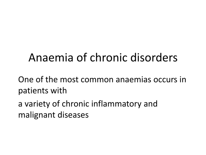
Anaemia of chronic disorders One of the most common anaemias occurs in patients with a variety of chronic inflammatory and malignant diseases
The characteristic features 1 Normochromic, normocytic or mildly hypochromic (MCV rarely <75fL) indices and red cell morphology. 2 Mild and non progressive anaemia (haemoglobin rarely <90 g/L) the severity being related to the severity of the disease. 3 Both the serum iron and TIBC are reduced. 4 The serum ferritin is normal or raised. 5 Bone marrow storage (reticuloendothelial) iron is normal but erythroblast iron is reduced
The pathogenesis decreased release of iron from macrophages to plasma because of raised serum hepcidin levels reduced red cell lifespan and an inadequate erythropoietin response to anaemia caused by the effects of cytokines such as IL 1 and tumour necrosis factor (TNF) on erythropoiesis.
The anaemia is corrected by successful treatment of the underlying disease and does not respond to iron therapy. Erythropoietin injections improve the anaemia in some cases. In many conditions this anaemia is complicated by anaemia resulting from other causes (e.g. iron, vitamin B12 or folate deficiency, renal failure, bone marrow failure, hypersplenism, endocrine abnormality leucoerythroblastic anaemia), discussed later
Sideroblastic anaemia This is a refractory anaemia defined by the presence of many pathological ring sideroblasts in the bone marrow These are abnormal erythroblasts containing numerous iron granules arranged in a ring or collar around the nucleus instead of the few randomly distributed iron granules seen when normal erythroblasts are stained for Iron There is also usually erythroid hyperplasia with ineffective erythropoiesis
Sideroblastic anaemia is diagnosed when 15% or more of marrow erythroblasts are ring sideroblasts. They can be found at lower numbers in a variety of haematological conditions.
hereditary forms the anaemia is usually characterized by a markedly hypochromic and microcytic blood picture. The most common mutations are in the ALA S gene which is on the X chromosome. Pyridoxal 6 phosphate is a coenzyme for ALA S. Other rare types include an X linked disease with spinocerebellar degeneration and ataxia, mitochondrial defects (e.g. Pearson s syndrome when there is also pancreatic insufficiency), thiamine responsive and other autosomal defects
In some patients, particularly with the hereditary type, there is a response to pyridoxine therapy. Folate deficiency may occur and folic acid therapy may also be tried. Other treatments, e.g. erythropoietin, may be tried in the myelodysplasia form In many severe cases, repeated blood transfusions are the only method of maintaining a satisfactory haemoglobin concentration
Lead poisoning Lead inhibits both haem and globin synthesis at a number of points. In addition it interferes with the breakdown of RNA by inhibiting the enzyme pyrimidine 5 nucleotidase, causing accumulation of denatured RNA in red cells, the RNA giving an appearance called basophilic stippling on the ordinary (Romanowsky) stain The anaemia may be hypochromic or predominantly haemolytic, and the bone marrow may show ring sideroblasts. Free erythrocyte protoporphyrin is raised.
Differential diagnosis of hypochromic anaemia The clinical history is particularly important as the source of the haemorrhage leading to iron deficiency or the presence of a chronic disease may be revealed. The country of origin and the family history may suggest a possible diagnosis of thalassaemia or other genetic defect of haemoglobin. Physical examination may also be helpful in determining a site of haemorrhage, features of a chronic inflammatory or malignant disease, koilonychia or, in some haemoglobinopathies, an enlarged spleen or bony deformities.
In thalassaemia trait the red cells tend to be small, often with an MCV of 70 fL or less, even when anaemia is mild or absent; the red cell count is usually over 5.5 1012/L. Conversely, in iron deficiency anaemia the indices fall progressively with the degree of anaemia and when anaemia is mild the indices are normal or only just reduced below normal (e.g. MCV 75 80 fL). In the anaemia of chronic disorders the indices are also not markedly low, an MCV in the range 75 82 fL being usual.
It is usual to perform a serum iron and TIBC measurement, or alternatively serum ferritin estimation, to confirm a diagnosis of iron deficiency. Haemoglobin high performance liquid chromatography (HPLC) or electrophoresis with an estimation of Hb A2 and Hb F is carried out in all patients suspected of thalassaemia or other genetic defect of haemoglobin
Thalassaemia trait is characterized by a raised Hb A2 above 3.5%, but in thalassaemia trait there is no abnormality on simple haemoglobin studies so the diagnosis is usually made by exclusion of all other causes of hypochromic red cells and by the presence of a red cell count >5.5 1012/L. DNA studies can be used to confirm the diagnosis.
Bone marrow examination is essential if a diagnosis of sideroblastic anaemia is suspected but is not usually needed in diagnosis of the other hypochromic anaemias.


