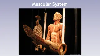
Anatomy of Muscles and Their Actions Illustrated
Explore the anatomy of key muscles like Gastrocnemius, Soleus, Tibialis Anterior, Peroneus Longus and Brevis, Deltoid, Pectoralis Major, and Trapezius. Learn about their origins, insertions, and actions in the human body with detailed images and descriptions.

Download Presentation

Please find below an Image/Link to download the presentation.
The content on the website is provided AS IS for your information and personal use only. It may not be sold, licensed, or shared on other websites without obtaining consent from the author. If you encounter any issues during the download, it is possible that the publisher has removed the file from their server.
You are allowed to download the files provided on this website for personal or commercial use, subject to the condition that they are used lawfully. All files are the property of their respective owners.
The content on the website is provided AS IS for your information and personal use only. It may not be sold, licensed, or shared on other websites without obtaining consent from the author.
E N D
Presentation Transcript
Gastrocnemius forms the most prominent contour of the calf. Origin: Medial Head: Lower medial condyle of femur. Lateral head: Lower lateral condyle of femur. Insertion : Posterior surface of Calcaneus via the calcaneal tendon. Action : Plantar flexion of foot and assists in flexion of Knee joint
Soleus Soleus lies beneath Gastrocnemius Origin: Upper posterior tibia and fibula Insertion With Gastrocnemius via Calcaneal tendon to calcaneus. Action Plantar flexion of the ankle joint.
Tibialis Anterior Origin = Lateral condyle of Tibia Insertion = Medial Cuneiform bone and base of 1st metatarsal Action =Dorsiflexion and inverts the foot.
Peroneus Longus and Brevis are part of the Peroneal muscle group. They lie on the lateral side of lower leg and evert the foot. Origin Peroneus Longus arises from head of fibula Peroneus Brevis arises from upper 2/3 of fibula Insertion Peroneus Longus: base of underside of 1st metatarsal and cuneiforms. Peroneus Brevis: base of 5th metatarsal Action Eversion Plantarflexion
Gastrocnemius Peroneus Brevis Soleus
Deltoid is a triangular shaped muscle and encapsulates the shoulder. spine of Scapula. Origin - Clavicle, Acromion process and Insertion - Deltoid Tuberosity of Humerus. Anterior fibres = Flexes and medially rotates Humerus. Middle fibres = Abducts Humerus. Posterior Fibres = Extends and Laterally rotates Humerus Action
Pectoralis Major - is a fan shaped muscle of the anterior chest. Origin Clavicular head Medial of clavicle. Sternocostal portion sternum and upper 6 costal cartilages. Insertion Upper shaft of Humerus. Action: Clavicular portion Flexes and medially rotates humerus. Horizontally adducts humerus to opposite shoulder (.applying deodorant). Sternocostal portion Adducts Humerus to opposite hip (Pull down from above).
The left and right Trapezius creates a trapezium in shape, giving its name. Origin: Base of Skull, Spinous processes of 7th Cervical vertebrae and all Thoracic vertebrae. Insertion: Lateral third of clavicle, Acromion process, Spine of scapula. Action: Upper fibres: elevates shoulder girdle Middle fibres: retract scapula. Lower fibres: Depress scapula.
Latissimus Dorsi also forms posterior wall of axilla. Origin: Lower 6 thoracic vertebrae, all lumbar and sacral vertebrae, posterior iliac crest, lower 3 or 4 ribs and inferior angle of scapula! Insertion: Bicipital groove at the top of Humerus. Action: Adducts and medially rotates humerus. Extends the flexed arm.