Autoimmune Hemolytic Anemia: Causes, Diagnosis, and Treatment
Discover the complexities of autoimmune hemolytic anemia (AIHA), its different forms, pathogenesis, and treatment options. Learn about warm AIHA, cold agglutinin disease, and more in this comprehensive review article. Gain insights into the immune mechanisms underlying AIHA and the latest findings in the field.

Download Presentation

Please find below an Image/Link to download the presentation.
The content on the website is provided AS IS for your information and personal use only. It may not be sold, licensed, or shared on other websites without obtaining consent from the author. If you encounter any issues during the download, it is possible that the publisher has removed the file from their server.
You are allowed to download the files provided on this website for personal or commercial use, subject to the condition that they are used lawfully. All files are the property of their respective owners.
The content on the website is provided AS IS for your information and personal use only. It may not be sold, licensed, or shared on other websites without obtaining consent from the author.
E N D
Presentation Transcript
JOURNAL CLUB REVIEW ARTICLE 06/04/2022 BIBYMOL JOSEPH JR2
AUTOIMMUNE HEMOLYTIC ANEMIAS SIGBJORN BERENTSTEN WILMA BARCELLINI
INTRODUCTION Autoimmune hemolytic anemia (AIHA) is defined as increased destruction of red cells through autoimmune mechanisms, usually mediated by autoantibodies against erythrocyte surface antigens. During the past decade, important new findings have emerged regarding the cause, pathogenesis, diagnosis, and treatment of this group of disorders. AIHA is a common term for several diseases that differ from one another with respect to cause, pathogenesis, and clinical presentation, and the individual disorders should be addressed according to these differences.
WARM AIHA COLD AGGLUTININ DISEASE SECONDARY COLD AGGLUTININ SYNDROME PAROXYSMAL COLD HAEMOGLOBINURUA MIXED AIHA DRUG INDUCED AIHA
WARM AIHA In warm AIHA,antibodies have the highest affinity to the antigen at 37 C. The autoantibodies are polyclonal (i.e., produced by nonclonal B lymphocytes and plasma cells). They are usually of the IgG class, but IgM warm antibodies may be involved, and in rare cases, IgA warm antibodies may be involved.
PATHOGENESIS The pathogenesis of warmAIHA is complex The mononuclear phagocytic system in the spleen plays a major role in the breakdown of opsonized erythrocytes (extravascular hemolysis). The complement system, activated by the classical pathway, is involved in approximately half the cases. Most of the complement-mediated erythrocyte destruction occurs through phagocytosis of complement fragment 3b (C3b) coated cells (extravascular hemolysis).
To a lesser extent, cleavage of C5 and activation of the terminal complement cascade may occur, resulting in formation of the membrane attack complex and intravascular hemolysis. The pathogenesis of this disorder also involves the T-lymphocyte system The signal substance cytotoxic T-lymphocyte antigen 4 (CTLA-4) activates regulatory T cells, which are critical for immune tolerance.
Polymorphism predisposition autoimmune cytopenias. The programmed cell death 1 (PD-1) signal pathway is another checkpoint for pharmacologic inhibition of PD-1 carries an increased risk of autoimmune disease. of to the immunologic CTLA-4 gene disorders, can confer including a immune tolerance, and
In nearly 50% of cases of warm AIHA, no underlying or associated disorder can be identified, and the hemolytic disease is classified as primary. Slightly more than 50% of the cases are secondary to immunologic or lymphoproliferative disorders (e.g., chronic lymphocytic erythematosus,or common variable immunodeficiency). leukemia, systemic lupus
Occasionally,a lymphoproliferative disease develops several years after the diagnosis ofAIHA. Recently, several cases have been described in patients with severe acute respiratory syndrome coronavirus 2 (SARS-CoV-2) infection. The Evans syndrome, characterized by the simultaneous or sequential combination of AIHA and immune thrombocytopenia, is often associated with more severe anemia and a worse prognosis than primary warmAIHA.
COLD AGGLUTININ DISEASE Cold-reactive antibodies bind to the antigen at a temperature of 0 to 4 C but may also react at higher temperatures. Cold agglutinins:Cold autoantibodies that can agglutinate erythrocytes. The thermal amplitude:the highest temperature at which agglutination can be detected, and cold agglutinins with a thermal amplitude higher than 28 to 30 C are pathogenic. The cold agglutinin titer: the inverse of the highest plasma or serum dilution at which agglutination can be seen at a given temperature.
Cold agglutinin disease is an AIHA in which the autoantibody is a cold agglutinin and no underlying clinical disorder is present. Newer studies have shown that affected patients, who previously would have received a diagnosis of primary or idiopathic cold agglutinin lymphoproliferative bone marrow disease that can be difficult to recognize. disease, have a clonal
The histopathological picture has often been interpreted as several types of low-grade nonHodgkin s lymphoma, such as lymphoplasmacytic or marginal-zone lymphoma, but a relatively uniform disease, cold agglutinin associated lymphoproliferative bone marrow disorder, is now thought to be the underlying condition The MYD88 L265P mutation, present in nearly all cases of lymphoplasmacytic lymphoma (the bone marrow disorder seen in Waldenstr m s macroglobulinemia), is usually not found in cold agglutinin disease.
In affected patients, the cold agglutinins are monoclonal, usually of the IgM class, and are produced by clonal lymphocytes in the bone marrow. Cooling of the blood in acral parts of the circulation allows for binding of cold agglutinin to its antigen on the erythrocyte surface, resulting in agglutination and impaired passage through the capillaries. Thus, about half of patients with cold agglutinin disease have cold-induced circulatory symptoms such as acrocyanosis or Raynaud-like phenomena. Gangrene is a rare complication.
PATHOGENESIS Hemolysis in cold agglutinin disease is complement-dependent. Activation of the classical pathway results in coating of erythrocytes with the split product C3b. C3b-opsonized cells are prone to phagocytosis by the mononuclear phagocytic system, (extravascular hemolysis). mainly in the liver
C3b can also react to form C5 convertase,which initiates the terminal complement cascade. Terminal complement activation, in turn, leads to formation of the membrane attack complex and intravascular hemolysis,at least in some patients and situations.
SECONDARY COLD AGGLUTININ SYNDROME The distinction between cold agglutinin disease and secondary cold agglutinin syndrome has been increasingly accepted. The latter is a rare, heterogeneous group of cold agglutinin- mediatedAIHA disorders that are secondary to other diseases - Mycoplasma pneumoniae infection Epstein Barr virus infection cytomegalovirus infection SARS-CoV-2 infection cancers (typically,aggressive B-cell lymphoma)
PAROXYSMAL COLD HEMOGLOBINURIA In paroxysmal cold hemoglobinuria, the autoantibody is a biphasic IgG hemolysin called Donath Landsteiner antibody. It binds to its antigen at temperatures below central body temperature, but activation of the classical complement pathway, beyond the initial steps, occurs after warming to 37 C in the central circulation. The terminal complement cascade is activated,and hemolysis is predominantly intravascular. Today, this disease occurs almost exclusively as a rare, temporary,post-viral complication in children.
MIXED AIHA Mixed warm antibody mediated and cold antibody mediated AIHA is defined by the presence of warm IgG autoantibodies combined with high-titer cold agglutinins. The mixed form of AIHA is often characterized by lower hemoglobin levels and a worse prognosis than warm AIHA, and two or more lines of therapy are frequently needed.
DRUG-INDUCED HEMOLYTIC ANEMIA Hemolytic anemia induced by drugs is usually mediated by immunologic mechanisms. More than 150 drugs have been implicated as causes of this condition,which nonetheless remains rare. Cases can be classified into two subtypes. In the drug-dependent subtype, autoantibodies are produced in response to a neoantigen formed by the binding of a drug to a cell-surface structure. In the drug-independent subtype, the offending drug can induce an autoimmune response that persists even in the absence of the drug.
Historically, methyldopa and large doses of penicillin were the most frequent causes of drug-induced hemolytic anemia. Today,most cases are caused by ceftriaxone other cephalosporins piperacillin nonsteroidal antiinflam matory drugs. Fludarabine, chlorambucil, and even bendamustine can increase the risk of autoimmune hemolysis when they are used to treat chronic lymphocytic leukemia,especially as monotherapy. checkpoint inhibitors that are used to treat cancer,in particular PD-1 inhibitors,can also induceAIHA.
AIHA AND THROMBOSIS WarmAIHA is associated with a risk of thrombosis. Three studies have reported thrombotic events in 11 to 20% of patients with AIHA, mostly deep venous thrombosis, with or without pulmonary embolism, but also venous thrombosis in atypical locations and arterial thromboembolism. Recent studies indicate an increased occurrence thrombosis even among patients with cold agglutinin disease, at a relative risk of 1.7 to 2.4 for affected patients as compared with the general population of
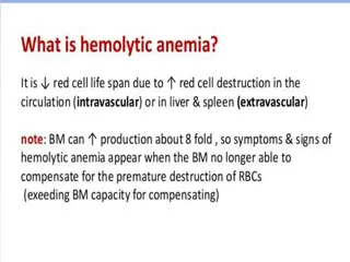
DIAGNOSIS Hemolytic anemia is diagnosed on the basis of the hemoglobin level and serum levels of lactate dehydrogenase, unconjugated bilirubin,and haptoglobin. Absolute reticulocytosis supports the presence of hemolysis, but the reticulocyte count may be normal or low, probably because of autoantibody precursors or a coexisting bone marrow disorder. activity against erythrocyte
Autoimmune pathogenesis is detected by means of the direct antiglobulin test (DAT,also termed Coombs test). A positive test shows immunoglobulin, complement, or both on the erythrocyte surface. A positive polyspecific (simple) DAT must be followed by a monospecific (extended) DAT, which immunoglobulin class (or classes) or complement protein bound to the red cell. Complement components, most often C3d, are detected in nearly half the cases of warm AIHA because of complement activation. will identify the
If warm-reactive IgM is involved, the DAT may be positive or negative for IgM because IgM may have detached from the cell before it can be detected in the laboratory. IgM is a potent complement activator and will leave the erythrocytes coated with C3. Therefore, a monospecific DAT that is positive for C3d and negative for IgG indicates IgM involvement.
The DAT is negative in 3 to 10% of patients with AIHA, depending on the DAT technique used. AIHA with a negative DAT remains a difficult diagnosis of exclusion. A false negative result can be due to a low density of immunoglobulin,complement,or both on the cell surface. A rare cause of false negative results is warm AIHA with IgA as the only autoantibody class, since most polyspecific reagents do not contain anti-IgA and thus cannot detect IgA.
This problem can be overcome by performing a monospecific DAT even when the polyspecific test is negative. More sensitive DAT methods (microcolumn, solid phase, cold washes, enzyme-linked immunosorbent assay, flow cytometry, dual DAT, and mitogen-stimulated laboratories may be useful. Nonimmune causes of hemolysis (congenital, toxic, or mechanical causes, as well as drugs and paroxysmal nocturnal hemoglobinuria) should be ruled out. DAT) in reference
In cold agglutinin disease and the cold agglutinin syndrome, the DAT is positive for C3d by definition and is usually negative for immunoglobulins but is weakly positive for IgG in up to 20% of cases. This result is not sufficient for establishing the diagnosis. The cold agglutinin titer must be assessed; it will be at least 64 (an integer that represents the activity of the antibody and the inverse of the dilution [1:64]) and usually much higher.
Paroxysmal cold hemoglobinuria should be suspected in children with acute hemolytic anemia and should be confirmed or ruled out with the Donath Landsteiner test. In a positive test, binding of the antibody to red cells occurs at a low temperature, followed by complement-mediated hemolysis on subsequent warming. Cold agglutinins are not present in this disease, and the DAT is frequently positive for C3 and negative for IgG.
Once warm AIHA has been diagnosed, an assessment for causes of secondaryAIHA should be undertaken. This includes a careful review of the medical history, a thorough clinical examination, assessment of serologic markers of autoimmune diseases,and relevant viral serologic tests. Whole-body computed tomography and flow cytometry in peripheral blood should be considered. Serum protein electrophoresis immunoglobulin classes must be performed for all types of AIHA. and quantification of
In cold agglutinin disease, the lymphoproliferative bone marrow disorder should be identified by biopsy and flow cytometry, although in a minority of cases, it will not be detected or interpretation of the results will be difficult. The relevant clonal bone marrow changes are most frequently identified in specialized pathology laboratories that are familiar with this entity. Even in patients with warm AIHA, bone marrow examinations should be performed for a wide range of indications,at least in case of therapy failure.
Cold agglutinins are sometimes incidentally detected in blood samples obtained for unrelated indications. In these cases, the patients often have a polyclonal, low-titer, low-thermal-amplitude cold agglutinin without hemolysis or clinical symptoms, and they do not have cold agglutinin disease or syndrome. Likewise, persons with incidental positive DAT results and no signs of hemolysis do not haveAIHA.
TREATMENT PRIMARYWARMAIHA Prednisolone (or prednisone) at a dose of 60 to 100 mg, or 1 mg per kilogram of body weight, per day is still recommended as initial first-line therapy for primary warmAIHA. After 2 to 3 weeks, dose tapering should be started in patients who have a response. prednisolone should be withdrawn if no response is observed.
If the dose can be reduced to 7.5 to 10 mg per day after 3 to 6 months without relapse anemia,further tapering should be attempted until the drug is discontinued. Approximately 80% of patients have an initial response to this regimen. However, only 30 to 40% have a sustained remission after 1 year. of clinically significant
The addition of rituximab to first-line therapy has been studied in two prospective, randomized trials. The results were almost identical, showing a doubling of long-term responses in the group that received both drugs.
The First International Consensus Group has recommended considering the addition of rituximab as initial therapy in patients with severe disease (i.e., those with a hemoglobin level of <8 g per deciliter). Atypical AIHA (IgA-mediated,mixed,or DAT-negative cases) and the Evans syndrome are also regarded as severe disease in this context. In patients who do not have a response to first-line therapy, a diagnostic reevaluation should be considered,focusing on any previously overlooked cause of secondaryAIHA.
The currently recommended second-line therapy for primary disease is rituximab,if it has not been added to first-line therapy. Response rates of 70 to 80% have been reported, with a median time to a response of 3 to 6 weeks. However,30% of patients have a relapse within 3 years The conventional dose is 375 mg per square meter of body- surface area, administered weekly for four cycles, or a 1000-mg fixed dose, administered for two cycles at a 2-week interval.
Splenectomy is currently recommended for patients who do not have a response to or who have a relapse after rituximab therapy. A response is seen in approximately 70% of patients, but the rate of long-lasting remission is unknown. The drawbacks of splenectomy are a risk of severe infection and a further increased risk of thrombosis. Other options for third-line or subsequent therapy are azathioprine, cyclophosphamide, cyclosporine, mycophenolate mofetil, and bortezomib.
In patients who initially have a response to glucocorticoids, long-term, low-dose prednisolone ( 10 mg daily) may be an appropriate approach. For emergencies, high-dose intravenous methylprednisolone, intravenous immune globulin, complement C1 inhibition, plasma exchange, splenectomy, and partial splenic embolization have been tried with some success in single patients or retrospective series.
SECONDARYWARMAIHA With some exceptions, first-line therapy for secondary warm AIHA should be the same as first line therapy for the primary form. Specific therapy for the underlying or associated disease is indicated if it requires treatment by itself or if the hemolytic anemia is resistant to first-line therapy [for example, in AIHA complicating chronic lymphocytic leukemia]
COLD AGGLUTININ DISEASE Glucocorticoids should not be used to treat cold agglutinin disease because response rates are low and patients who have a response often require unacceptably high doses to maintain the remission. Patients with mild anemia or compensated hemolysis should be monitored without treatment if circulatory symptoms and fatigue are absent or tolerable. Nonpharmacologic management: consists of thermal protection to limit hemolysis and relieve any ischemic symptoms.
Among treatments directed at the pathogenic B-cell clone, rituximab monotherapy (four doses of 375 mg per square meter at 1-week intervals) has become the most commonly used first- line therapy for cold agglutinin disease. The addition of fludarabine has yielded much higher overall and complete response rates but also more acute and late-onset toxic effects.