Cirrhosis: Causes, Types, and Risk Factors
Chronic liver disease characterized by scarring of the liver, leading to impaired liver function. Common causes include chronic viral infections, alcohol abuse, and obesity. Different types of cirrhosis exist, such as alcoholic, biliary, and postnecrotic cirrhosis. Risk factors include excessive alcohol consumption, obesity, and viral hepatitis. Identifying the causes, types, and risk factors of cirrhosis is crucial for prevention and management of this serious condition, which is a leading cause of death in India.

Download Presentation

Please find below an Image/Link to download the presentation.
The content on the website is provided AS IS for your information and personal use only. It may not be sold, licensed, or shared on other websites without obtaining consent from the author.If you encounter any issues during the download, it is possible that the publisher has removed the file from their server.
You are allowed to download the files provided on this website for personal or commercial use, subject to the condition that they are used lawfully. All files are the property of their respective owners.
The content on the website is provided AS IS for your information and personal use only. It may not be sold, licensed, or shared on other websites without obtaining consent from the author.
E N D
Presentation Transcript
MR. SHAILENDRA VERMA NURSING TUTOR RCN, BAREILLY
INTRODUCTION The word "cirrhosis" is a neologism that derives from Greek kirrhos, meaning "tawny" (the orange-yellow colourof the diseased liver). The destruction of the normal structure and the loss of liver cells prevent the liver from functioning normally
DEFINITION It is a chronic liver disease characterized by scarring of the liver that involves the formation of fibrous (scar) tissue associated with the destruction of the normal parenchymal cells of the organ.
INCIDENCE Around 10 lakh patients of liver cirrhosis are newly diagnosed every year in India. - Liver disease is the tenth most common cause of death in India as per the World Health Organization.
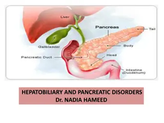
TYPES Alcoholic cirrhosis- Most common, due to chronic alcoholism. Scar tissue characteristically surrounds the portal area. Postnecroticcirrhosis- There is broad bands of scar tissue due to late results of acute viral hepatitis, postintoxicationwith industrial chemicals. Biliarycirrhosis- Scaring occurs around bile duct in liver, Results from chronic biliary obstruction & infection. Cardiac cirrhosis (right sided heart failure) and Nonspecific metabolic cirrhosis (GI diseases).
CAUSES The most common causes of cirrhosis are chronic (long-term) viral infections of the liver (hepatitis types B, and C), fatty liverassociated with obesityand diabetes, and alcohol abuse. Chronic abuse of alcohol (drinking too much alcohol for a long time) Chronic hepatitis C (hepatitis C that has lasted at least 6 months) Alpha-1 antitrypsin deficiency hemochromatosis Wilson's disease Blockage of the bile duct.
RISK FACTORS Drinking too much alcohol. Being overweight. Having viral hepatitis.
PATHOPHYSIOLOGY Primary event is injury to hepatocellularelements Initiates inflammatory response with cytokine release- >toxic substances Destruction of hepatocytes, bile duct cells, vascular endothelial cells Repair through cellular proliferation and regeneration Formation of fibrous scar CIRRHOSIS OF LIVER
SIGN AND SYMPTOMS Loss of appetite Lack of energy (fatigue) Weight loss or sudden weight gain Bruises Yellowing of skin or the whites of eyes (jaundice) Itchy skin Fluid retention (edema) and swelling in the ankles, legs, and abdomen A brownish or orange color to the urine Confusion, disorientation, personality changes Blood in the stool Fever
DAIGNOSTIC EVALUATION History and Physical examination Blood tests: computerized tomography (CT) USG Biopsy: Surgery: In some cases, cirrhosis is diagnosed during surgery when the doctor is able to see the entire liver. The liver also can be inspected through a laparoscope, a viewing device that is inserted through a tiny incision (cut) in the abdomen
COMPLICATION Kidney failure Reduced oxygen in the blood Diabetes Changes in blood counts Increased risk of infections Excessive bleeding and bruising Breast enlargement in men Premature menopause Variceal bleeding DEATH
MANAGEMENT 1. NON-SURGICAL Traditionally, liver damage from cirrhosis cannot be reversed, but treatment could not stop or delay further progression and reduce complications. Close follow-up is often necessary. Antibiotics will be prescribed for infections. Treating underlyng cause Alcoholic cirrhosis caused by alcohol abuse is treated by abstaining from alcohol. Treatment for hepatitis-related cirrhosis involves medications used to treat the different types of hepatitis, such as interferon for viral hepatitis. Treatment of cirrhosis includes 1) preventing further damage to the liver, 2) treating the complications of cirrhosis, 3) preventing liver cancer or detecting it early.
1) preventing further damage to the liver Consume a balanced diet and one multivitamin daily. Avoid drugs (including alcohol) that cause liver damage. All patients with cirrhosis should avoid alcohol. Avoid nonsteroidal antiinflammatorydrugs (NSAIDs, e.g. ibuprofen). Eradicate hepatitis B and hepatitis C virus by using anti-viral medications.
2) preventing liver cancer or early detecting Several types of liver disease that cause cirrhosis are associated with a particularly high incidence of liver cancer, for example, hepatitis B and C, and it would be useful to screen for liver cancer since early surgical treatment or transplantation of the liver can cure the patient of cancer.
3) Treating the complications of cirrhosis Edema and ascites. Retention of salt and water can lead to swelling of the ankles and legs (edema) or abdomen (ascites) in patients with cirrhosis. Doctors often advise patients with cirrhosis to restrict dietary salt (sodium) and fluid to decrease edema and ascites. Diuretics are medications that work in the kidneys to promote the elimination of salt and water into the urine. A combination of the diuretics spironolactone (Aldactone) and furosemidecan reduce or eliminate the edema and ascites in most patients. It is common to withdraw large amounts (liters) of fluid from the abdomen when the ascites is causing painful abdominal distension
Bleeding from verices Propranolol (Inderal), a beta blocker, is effective in lowering pressure in the portal vein and is used to prevent initial bleeding and rebleeding from varices in patients with cirrhosis. Nitrates often are added to propranolol if propranolol alone does not adequately lower portal pressure or prevent bleeding. Octreotide (Sandostatin) also decreases portal vein pressure and has been used to treat variceal bleeding. During upper endoscopy (EGD), either sclerotherapy or band ligation can be performed to obliterate varices and stop active bleeding and prevent rebleeding
SURGICAL MANAGEMENT Band Ligation: - Endoscopy, which consists of a narrow tube mounted with a video camera being inserted into the throat, is used to identify the bleeding site. A rubber band is used to tie off the bleeding portion of the vein. TransjugularIntrahepatic Portosystemic Shunting (TIPS): - This procedure is done under 2 conditions: 1. Abdominal fluid which does not respond to standard medical treatment 2. Acute bleeding from esophageal varices unresponsive to standard medical or endoscopic treatment
Other Surgical Shunts: - The splenorenal shunt: - helps to reduce the pressure within the variceal system by connecting the spleen vein to a kidney vein. portacaval shunt: - reduces pressure in the entire portal system by connecting the portal vein to the inferior vena cava. Paracentesis It is simply takes fluid out from the abdominal cavity. In this procedure, a soft catheter is inserted into the abdomen. Usually when large volumes of fluid are to be removed, human albumin is introduced into the abdominal cavity. LIVER TRANSPLANTATION
Acute pain related to liver enlargement secondary to ascitesas evidenced by facial grimace. Activity intolerance related to fatigue and discomfort. Risk for injury and bleeding related to altered clotting mechanisms. Fluid volume excess related to ascitesand edema formation. Imbalanced nutritional status, less than body requirements, related to less intake of food Altered breathing pattern related to decreased lung expansion secondary to intra-abdominal fluid collection (ascites)
ASSIGNMENT :- Assignment on NCP of pre and post surgical management. Date of Submission ---------------


 preventing further damage to")
 preventing liver cancer or early")
 Treating the complications of")