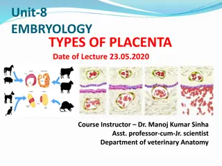
Development of Placenta in Embryology
The placenta is a crucial structure formed during pregnancy for the exchange of nutrients and waste materials between the embryo and mother. It develops from the chorio-allantoic membrane of the fetus and the maternal endometrium. The process begins with blastocyst implantation, leading to the transformation of the endometrium into decidua. The decidua consists of distinct layers serving different functions. Implantation of the conceptus in the endometrium occurs through phases of apposition, adhesion, and attachment, allowing for proper nourishment of the embryo. The gradual process of implantation varies among different animal species.

Download Presentation

Please find below an Image/Link to download the presentation.
The content on the website is provided AS IS for your information and personal use only. It may not be sold, licensed, or shared on other websites without obtaining consent from the author.If you encounter any issues during the download, it is possible that the publisher has removed the file from their server.
You are allowed to download the files provided on this website for personal or commercial use, subject to the condition that they are used lawfully. All files are the property of their respective owners.
The content on the website is provided AS IS for your information and personal use only. It may not be sold, licensed, or shared on other websites without obtaining consent from the author.
E N D
Presentation Transcript
VAN-608 EMBRYOLOGY Topic - General embryology (Implantation of Embryo & Development of Placenta) Course Instructor Dr. Manoj Kumar Sinha Department of Veterinary Anatomy
DEVELOPMENT OF PLACENTA Recall the previous classes and start from zygote formation : to the placenta formation
DEVELOPMENT OF PLACENTA The placenta is a foetomaternal composite structure formed by the association of embryo and extra embryonic membrane with uterine tissue for exchange of food materials , oxygen and waste materials Placenta develops from two sources: Foetal part From chorio-allantoic membrane Maternal part From Endometrium (decidua basils) Placenta begins to develop upon implantation of the blastocyst into the maternal endometrium (That means development of placenta starts when blastocyst attached to the endometrium) Once blastocyst is embeded endometrium changed into Decidua and secretory activity of endometrium started, glycogen and lipids are stored and vacuole appear into the stroma Placenta grows throughout the pregnancy in the endometrial wall,
Decidua Decidua (cells loaded with lipid and glycogen) is the term for the uterine lining during a pregnancy (endometrium changes into which is more vascular and more fuctional called decidua) Three layer: 1. Decidua basils : where the implantation takes place and the basal plate is formed 2. Decidua capsularis : lies like a capsule around chorion 3. Decidua Parietalis/vera : on the opposite uterus wall (endometrium) 1. functional part 3. 2.
Implantation The embryo along with extra-embryonic membranes are called Conceptus and attachment of conceptus to the endometrium is Implantation Implantation occur in three phases: Apposition Adhesion and Attachment Embryo got nourishment in uterine tube by its own yolk and secretion from oviducts In uterus embryo derive nutrition from uterine fluid , uterine fluid consists of cellular debries, extravasated poly- morphonucleocutes and secretion of endometrial gland called uterine milk (histotrophs) Implantation is slow and gradual process in domestic animals There is marked species difference in time of implantation, gestation period and litter size
Time of implantation Gestation period (in days) Species Litter size Cow 28-35 282 (277-290) 01 Ewe 17-20 148 (144-152) 1-2 Sow 17-24 114 (110-116) 08-12 Mare 49-70 338 (330-345) 01 Bitch 14-21 61 (58-64) Multiple Cat 14-21 64 (60-68) 04
Types of Implantation Three types of Implantation: Superficial/Central: chorionic within uterine cavity and expands to fill its lumen Ex. Domestic Animals Ecentric : vesicle become embeded in pockets of the uterine wall Ex. Rat , Squirrel Interstitial : The blastocyst penetrate into the wall of uterus and develops there until parturition Ex. Primates The 1. vesicle remain The chorionic partially 2. 3. 1. 2. 3.
Continue.. At the time of implantation Zona pellucida becomes disappear The trophoblastic layer differentiates into two parts: Inner layer - Cytotrophoblast Outer layer - Syncytotrophoblast Syncytotrophoblast proliferates into multilayered, multinucleated protoplasmic mass Cytotrophoblast differentiates into layer of primary mesoderm Primary mesoderm Syncytotrophoblast Cytotophoblast CHORION
Continue.. Inside syncytotrophoblast a number of lacunar spaces appear and syncytial cells form cords between the lacunar space, called Trabeclae Cords of cytotrophoblast invade the trabeculae and convert into Primary chorionic villi , lacunar space are now called intervillous space Primary chorionic villi are transformed into Secondary chorionic villi when primary mesodermic layer invade into the primary villi Secondary villi are transformed into Tertiary villi when the foetal blood vessels appear within primary mesoderm and their branches project into secondary villi Later on within primary mesoderm vacuoles are appeared subsequently they coalase to form extra-embryonic coelome between amniotic cavity and primary mesoderm
Continue In Birds and in some Farm animals like Cattle, Sheep, Goat and Pig the allantoic vesicle expands into the extroembryonic coelom and surrounds the whole amniotic cavity It occupies the space between the amnion and the chorion (serosa), the outer wall of amnion fuses with chorion and therefore forms Chorioallantioic Type of Placenta The amniotic cavity contains Amniotic fluids within which embryo becomes float. Amniotic fluid contains salt, water, protein and sugar . It gives protection to the foetus by neutralizing shock and pressure. It also acts as lubricants at the time of birth At the time of birth the placenta is discarded along with the amnion and referred as Afterbirth
Continue In Bovines, attachment between maternal and foetal membranes occur throughout the endometrium of the horn in the sporadic manner as per distribution of cotyledones In Sow and Mare the union of chorion and uterine wall is superficial and their separation at the time of birth without injury to maternal tissue this type of placenta is called Deciduate placenta In Carnivores The villi occupy on the girdle like band around the middle of chorionic sac In Humans, The chorionic villi develop rapidly at the embryonic pole of blastocyst called chorionic frondosum In Primates The union between foetal and maternal tissue is so intimate and damage of uterine tissue at the time of birth, that's why there is extensive bleeding at the time of birth in primates
Continue Exchange of metabolites occurs directly through foetal and maternal blood circulation There is no direct mixing of foetal and maternal blood in placenta The chorio-allantoic placenta directly absorb nutrition from maternal blood is called Haemotrope Therefore placenta formation (contact between foetal membrane and endometrium) occurs in various zones which differs characteristically depending upon the species Types of placenta in next class