Endocrine Disorders and Cardiovascular Disease: Insights from Dr. Vivek C. Warrier at GMC Calicut
Explore the intricate connection between the endocrine system and cardiovascular diseases as explained by Dr. Vivek C. Warrier, Senior Resident in the Department of Cardiology at GMC Calicut. Learn how hormones influence lipid metabolism and impact cardiac health, with a focus on pituitary hormones and growth hormone abnormalities leading to conditions like acromegaly and associated cardiovascular manifestations.

Download Presentation

Please find below an Image/Link to download the presentation.
The content on the website is provided AS IS for your information and personal use only. It may not be sold, licensed, or shared on other websites without obtaining consent from the author. If you encounter any issues during the download, it is possible that the publisher has removed the file from their server.
You are allowed to download the files provided on this website for personal or commercial use, subject to the condition that they are used lawfully. All files are the property of their respective owners.
The content on the website is provided AS IS for your information and personal use only. It may not be sold, licensed, or shared on other websites without obtaining consent from the author.
E N D
Presentation Transcript
Endocrine Disorders and Cardiovascular Disease DR VIVEK C WARRIER SENIOR RESIDENT DEPARTMENT OF CARDIOLOGY, GMC CALICUT
The endocrine system is linked closely with many important cardiovascular diseases. Hormones can alter the cardiovascular system through changes in lipid metabolism & actions on cardiac myocytes, vascular smooth muscle cells, & other target cells & tissues.
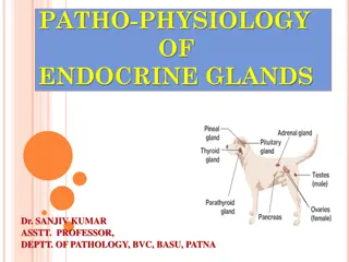
PITUITARY HORMONES ADRENAL HORMONES THYROID HORMONES PARATHYROID HORMONES
Pituitary Hormones and Cardiovascular Disease The anterior pituitary, or adenohypophysis, contains six different cell types; five of them produce polypeptide or glycoprotein hormones, & one, the sixth, consists of nonsecretory chromophobic cells. Of these cell types, the somatotropic cells, which secrete human growth hormone (hGH), & the corticotropic cells, which produce adrenocorticotropic hormone (ACTH), can contribute to cardiac disease.
Growth Hormone Excessive secretion of hGH & insulin-like growth factor type 1 (IGF-1) by benign pituitary adenomas leads to the clinical syndrome of gigantism in youth, & to acromegaly in adults. hGH exerts its cellular effects through two major pathways : Firstly, by binding of the hormone to specific hGH receptors on target cells. Such receptors exist in the heart, skeletal muscle, fat, liver, & kidneys, as well as in many additional cell types throughout fetal development. Secondly - stimulation of the synthesis of IGF-1. The liver produces most of the IGF-1, but other cell types can produce IGF-1 under the influence of hGH.
Cardiovascular Manifestations of Acromegaly About 60% of acromegalic patients develop cardiovascular disease. Hypertension, insulin resistance, diabetes mellitus, & hyperlipidemia are the cardiovascular risk factors associated most frequently with acromegaly. Death in acromegalic patients occurs primarily from cardiovascular disease & diabetes, especially in undiagnosed & untreated patients. The recently published Endocrine Society Clinical Practice Guidelines recommend that acromegalic patients undergo evaluation for associated comorbidities (hypertension, diabetes mellitus, cardiovascular disease, & sleep apnea).
The cardiovascular & hemodynamic effects of acromegaly vary considerably depending on the patient's age & the disease's severity & duration. A specific acromegalic cardiomyopathy develops in patients with persistently increased secretion of hGH & IGF-1. It is manifested as biventricular concentric hypertrophy. Two thirds of patients with acromegaly meet echocardiographic criteria for left ventricular hypertrophy (LVH); the right ventricular mass also increases in acromegaly, a finding indicating a more generalized process than systemic hypertension.
This specific cardiomyopathy has three phases. The first phase typically develops in young patients with new- onset acromegaly & involves a hyperkinetic syndrome with increased myocardial contractility & enhanced cardiac output. The second phase of cardiomyopathy has more evident hypertrophy & is associated with impaired diastolic filling, which reduces the cardiac performance during exercise. Third phase- Impaired systolic function & low cardiac output progressively develop in the late phase of the disease in patients in whom acromegaly is undiagnosed or undertreated. Heart failure can complicate this late phase of the disease
The clinical disease activity of patients with an excess of hGH is correlated better with serum levels of IGF-1 than with hGH concentrations. Secondary hypertension accompanies acromegaly & occurs with a mean prevalence of 33% to 46%. The mechanism underlying hypertension in acromegaly remains poorly understood. Administration of hGH promotes sodium retention & volume expansion, & IGF-1 has a potent antinatriuretic effect independent of any effect on aldosterone.
Studies of the renin-angiotensin aldosterone system have shown failure to inhibit release of renin optimally by volume expansion. Impaired glucose tolerance & diabetes mellitus occur in 15% to 38% of acromegalic patients. Acromegaly increases the prevalence of aortic & mitral valve disease, which persists despite cure of the acromegaly. Patients with acromegaly may exhibit dilation of the aortic root. Abnormalities on the electrocardiogram (ECG), including left axis deviation, septal Q waves, ST-T wave depression, abnormal QT dispersion, & conduction system defects, develop in up to 50% of patients with acromegaly.
Diagnosis In 99% of cases, acromegaly arises from benign adenomas of the anterior pituitary gland. At diagnosis most of these neoplasms are classified as macroadenomas (> 10 mm). The biochemical diagnosis of acromegaly depends on demonstrating elevated serum IGF-1 levels & the lack of suppression of hGH to less than 1 g/L following an oral glucose load. Localization of the tumor uses magnetic resonance imaging (MRI) of the pituitary gland.
Treatment Transsphenoidal surgery with resection of the adenoma cures 50% to 70% of patients. Preoperative medical therapy with somatostatin receptor agonist is recommended to reduce the surgical risk in patients with heart failure or severe comorbidities. The cardiovascular complications of acromegaly usually improve with treatment, & survival rates improve significantly in patients achieving disease remission as defined by the normalization of serum IGF-1 & serum hGH levels to less than 1 g/L. A residual tumor mass following surgery may require radiotherapy if medical therapy is unavailable, unsuccessful, or not tolerated.
Cardiovascular Manifestations of Growth Hormone Deficiency Untreated adult hGH-deficient patients can have cardiac & endothelial dysfunction, insulin resistance, a deranged lipid profile, increased carotid intima-media thickness, elevated inflammatory markers, increased body fat with abdominal obesity, hypercoagulability, & decreased skeletal muscle mass & strength. Early premature atherosclerosis can develop in hypopituitary patients not receiving hGH therapy.
Adrenal Hormones and Cardiovascular Disease Cushing Disease & Cushing Syndrome Cushing syndrome results from prolonged & inappropriately high exposure of tissues to glucocorticoids. Excessive cortisol secretion & its attendant clinical disease states can arise from excessive release of ACTH by the pituitary (Cushing disease) or through the adenomatous or rarely malignant neoplastic process arising in the adrenal gland itself (Cushing syndrome).
Clinical signs & symptoms of Cushing syndrome often develop in patients treated with exogenous steroids at doses equivalent to 20 mg of prednisone daily for more than 1 month. Several cardiac genes contain glucocorticoid response elements in their promoter regions that confer transcriptional-level glucocorticoid responsiveness. Include those that encode voltage-gated potassium channels, as well as protein kinases, which serve to phosphorylate & regulate the voltage-gated sodium channels. In addition, there are more rapidly acting, nontranscriptional pathways by which cortisol may regulate the activity of voltage-gated potassium channels.
The cardiac effects of Cushing syndrome arise from the effects of glucocorticoids on the heart, liver, skeletal muscle, & fat tissue. LVH & concentric remodeling can result. Glucocorticoid excess is also associated with left ventricular dysfunction, myocardial fibrosis, & dilated cardiomyopathy. Chronic cortisol hypersecretion causes central obesity, hypertension, insulin resistance, dyslipidemia, a prothrombotic state, & the metabolic syndrome.
Electrocardiographic changes of Cushing disease : The duration of the PR interval appears to be correlated inversely with adrenal cortisol production rates. The mechanism underlying this correlation may be related to expression or regulation of the voltage-gated sodium channel (SCN5A). Changes on the ECG, specifically in the PR & QT intervals, may also arise from the direct (nongenomic) effects of glucocorticoids on the voltage-gated potassium channel (Kv1.5) in excitable tissues.
A particular complex of cardiac & adrenal lesions, referred to as the Carney complex, is a combination of Cushing syndrome, cardiac myxoma, & a variety of pigmented dermal lesions. This monogenic autosomal dominant trait has been mapped to the q2 region of chromosome 17. Myxomas most commonly occur in the left atrium but can arise throughout the heart, can develop at young ages, & can be multicentric.
Diagnosis The diagnosis of Cushing disease & Cushing syndrome requires the demonstration of increased cortisol production as reflected by an elevated 24-hour urinary free cortisol level or nocturnal salivary cortisol level.
Treatment Initial resection of primary lesion(s) is recommended for underlying Cushing disease (based in the pituitary) & also for Cushing disease related to ectopic & adrenal causes. Transsphenoidal selective resection with or without postoperative radiation therapy can partially or completely reverse the increased ACTH. Cushing syndrome requires surgical removal of one adrenal gland (adrenal adenoma, adrenal carcinoma) or both adrenal glands (multiple nodular disease).
Immediately after surgery, cortisol & mineralocorticoid (fludrocortisone) need to be replaced to prevent adrenal insufficiency. Drug therapy before or after surgery can help control persistent cortisol production. Pasireotide (somatostatin analogue ) can decrease ACTH production from a pituitary tumor. The adrenal enzyme inhibitor ketoconazole may be used alone or in combination with metyrapone to enhance control of severe hypercortisolemia.
Mitotane is used primarily to treat adrenal carcinoma. Mifepristone blocks the direct effect of cortisol on tissues & leads to an improvement in hypertension &/or diabetes in 40% to 60% of patients.
Primary Hyperaldosteronism Aldosterone production by the zona glomerulosa is responsive to the renin-angiotensin system. Renin secretion responds primarily to changes in intravascular volume. Aldosterone synthesis & secretion depend primarily on regulation by angiotensin II, which binds to the angiotensin II type I receptor on cells of the zona glomerulosa.
Common causes of PA include an adrenal adenoma, unilateral or bilateral adrenal hyperplasia, or, in rare cases, an adrenal carcinoma or an inherited condition known as glucocorticoid- remediable aldosteronism. Aldosterone enters cells & binds to the mineralocorticoid receptor, which then is translocated to the nucleus & promotes the expression of aldosterone-responsive genes. Primary aldosteronism can induce development of cardiac hypertrophy, myocardial fibrosis, & diastolic dysfunction. More than 10% of hypertensive patients have primary aldosteronism, & normokalemic hypertension constitutes the most common presentation of the disease.
Primary aldosteronism is associated with higher rates of cardiovascular morbidity & mortality than age- & sex-matched patients with essential hypertension. Primary aldosteronism should be investigated in patients with (1) severe hypertension, (2) treatment-resistant hypertension, (3) hypertension with spontaneous or diuretic- induced hypokalemia, (4) hypertension with adrenal incidentaloma, (5) hypertension & sleep apnea, or (6) a family history of early-onset hypertension or cerebrovascular accident at a young age (< 40 years of age).
The plasma aldosterone/renin ratio detects possible PA.( ratio >20 , PAC > 15ng/dl) Mineralocorticoid receptor antagonists should be withdrawn for at least 4 weeks before testing. Patients with an abnormal aldosterone/renin ratio undergo one or more confirmatory tests to definitively confirm or exclude the diagnosis. After sodium loading, plasma aldosterone levels lower than 5 ng/dL make the diagnosis of PA unlikely. Levels above 10 ng/dL indicate very probable PA. Patients with spontaneous hypokalemia, plasma renin levels below detection levels, & plasma aldosterone concentrations of more than 20 ng/dL do not require further testing.
Treatment Patients with primary hyperaldosteronism & hypokalemia should receive slow-release potassium chloride supplementation to maintain plasma potassium. The aldosterone antagonist spironolactone or eplerenone (as a second choice) should be used to control hypertension, hypokalemia, & the deleterious cardiovascular effects of aldosterone hypersecretion.
Unilateral laparoscopic adrenalectomy can cure hypokalemia & improve or cure hypertension in patients with unilateral disease Patients with bilateral disease & those reluctant to undergo surgery should receive medical treatment with mineralocorticoid receptor antagonists. Genetic testing for familial hyperaldosteronism should be performed in patients with a family history of hypertension & stroke at a young age (< 40 years).
Addison Disease Primary adrenal insufficiency occurs when the adrenal cortex cannot produce sufficient glucocorticoids &/or mineralocorticoids. Acute Addisonian crisis, one of the most severe endocrine emergencies, is characterized by hypovolemia, hypotension, & acute cardiovascular collapse resulting from renal sodium wasting, hyperkalemia, & loss of vascular tone. Primary adrenal insufficiency arises most commonly from bilateral loss of adrenal function on an autoimmune basis; as a result of infection, hemorrhage, or metastatic malignancy.
Addison disease may be associated with other autoimmune disorders (e.g., Hashimoto thyroiditis, type 1 diabetes mellitus, autoimmune gastritis/pernicious anemia, & vitiligo). Secondary adrenal insufficiency, which results from pituitary- dependent loss of ACTH secretion, leads to a fall in glucocorticoid production; mineralocorticoid production remains at relatively normal levels. The noncardiac symptoms, including increased pigmentation, abdominal pain with nausea & vomiting, hypoglycemia, & weight loss, can be chronic; tachycardia, hypotension, hyponatremia, hyperkalemia, loss of autonomic tone, & cardiovascular collapse & crisis may develop, especially in acutely ill or untreated patients with Addison disease.
Blood pressure measurements uniformly show a low diastolic pressure (< 60 mm Hg) along with orthostatic changes that reflect loss of volume & acquired autonomic dysfunction. Laboratory findings (hyponatremia & hyperkalemia) indicate loss of aldosterone production (renin levels are high). Cardiac atrophy is an unusual condition; it is seen with malnutrition caused by anorexia, in astronauts after prolonged space flight, in populations with sodium-deficient diets, & characteristically with Addison disease (teardrop heart). This atrophy reflects a response to decreases in the cardiac workload because restoration of normal plasma volume with mineralocorticoid & glucocorticoid replacement increases ventricular mass.
Diagnosis Acute adrenal insufficiency characteristically occurs in the setting of acute stress, infection, or trauma in a patient with chronic autoimmune adrenal insufficiency. Acute adrenal insufficiency can develop in patients treated over a long term with suppressive doses of corticosteroids (> 10 mg of prednisone for > 1 month) if treatment is stopped precipitously or if an acute, severe, non endocrine-related illness arises. The diagnostic criteria include low cortisol levels (morning cortisol < 140 nmol/L [< 5 g/dL]) or cortisol levels that fail to rise above 500 nmol/L (20 g/dL) 30 or 60 minutes after an intravenous injection of 250 g of corticotropin.
Treatment Management of acute addisonian crisis requires adequate hydrocortisone replacement therapy (100 mg given as an initial intravenous bolus; then 100 mg every 8 to 12 hours for the first 24 hours, & tapering of the dose over the next 72 to 96 hours). Large volumes of normal saline can address the intravascular fluid deficit. Potential underlying precipitating causes (including infection, acute cardiac or cerebral ischemia, or intraabdominal emergency) require identification & treatment.
Long-term treatment of adrenal insufficiency consists of oral corticosteroid therapy (hydrocortisone 20 mg in two divided oral doses per day, or prednisone 5 mg administered orally once or twice daily). Patients with confirmed aldosterone deficiency should receive mineralocorticoid replacement with fluorohydrocortisone (starting dose, 50 to 100 g in adults).
Pheochromocytoma Pheochromocytomas are primarily benign tumors arising from neuroectodermal chromaffin cells; they usually arise in the adrenal medulla & abdomen, but they may arise anywhere in the plexus of sympathetic adrenergic nerves. About 20% are familial. Six different familial autosomal dominant diseases can be suspected clinically: neurofibromatosis type 1, multiple endocrine neoplasia type 2 (MEN2), von Hippel-Lindau syndrome, renal cell carcinoma with SDHB mutation, Carney triad (paragangliomas, gastric stromal tumors, pulmonary chondromas), & Carney-Stratakis syndrome (paragangliomas & gastric stromal sarcomas).
Clinical manifestations of pheochromocytoma include headache, palpitations, excessive sweating, tremulousness, chest pain, weight loss, & a variety of other constitutional complaints. Hypertension may be episodic but is usually constant & is paradoxically associated with orthostatic hypotension on arising in the morning. The paroxysmal attacks & classic symptoms result from episodic excessive catecholamine secretion. Hypertension caused by pheochromocytoma can first present at the time of elective surgical intervention for an unrelated condition.
As a result of release of norepinephrine with an increase in systemic vascular resistance, cardiac output is minimally increased despite increases in the heart rate. The ECG can show LVH, repolarization abnormalities, findings suggesting left ventricular strain. Impaired left ventricular function & cardiomyopathy have occurred in patients with pheochromocytoma. mechanisms include an increased left ventricular work & LVH from associated hypertension; potential adverse effects of excess catecholamines on myocyte structure & contractility; & changes in coronary arteries, including thickening of the media, which presumably impairs blood flow to the myocardium.
Reversible dilated hypertrophic cardiomyopathy & takotsubo cardiomyopathy are well-established cardiac manifestations of pheochromocytoma. The primary catecholamine released from adrenal pheochromocytomas is norepinephrine. Demonstration of elevated serum dopamine levels implies malignant transformation, which in turn suggests that the tumor may have arisen in an extraadrenal site. Rarely, pheochromocytoma can arise within the heart, presumably from chromaffin cells, which are part of the adrenergic autonomic paraganglia.
Diagnosis Quantitative 24-hour urinary fractionated metanephrine levels are the most reliable screening procedures; they provide a sensitivity of 97% & a specificity of 91%. CT is the first-choice imaging modality because of its excellent spatial resolution for the thorax, abdomen, & pelvis. MRI is recommended in patients with metastatic disease & for detection of skull base & neck paragangliomas.
Treatment Definitive treatment of pheochromocytoma requires removal of the lesion. Accurate preoperative localization reduces the operative mortality rate & eliminates the need for exploratory laparotomy. Endoscopic procedures are now standard for small tumors, & open resection is indicated for large tumors (e.g., > 6 cm) or invasive pheochromocytomas. Preoperative pharmacologic treatment includes 7 to 14 days of alpha-adrenergic blockade (usually with doxazosin, prazosin, or phenoxybenzamine) to normalize the blood pressure.
Beta-blocking drugs can normalize the heart rate but should follow the establishment of a sufficient alpha blockade. Before surgical treatment, a high sodium diet & fluid intake should be started to improve the blood volume contraction & prevent severe hypotension after tumor removal. Lifelong annual biochemical testing to assess for recurrent or metastatic disease is necessary.
Parathyroid Hormones and Cardiovascular Disease Diseases of the parathyroid glands can produce cardiovascular disease & alter cardiac function through two mechanisms. Parathyroid hormone (PTH) is a protein hormone that can affect the heart, vascular smooth muscle cells, & endothelial cells. PTH-induced changes in serum calcium levels also affect the cardiovascular system.
PTH can bind to its receptor and alter the spontaneous beating rate of neonatal cardiac myocytes through an increase in intracellular cyclic adenosine monophosphate (cAMP). PTH can also alter the calcium influx & cardiac contractility in adult cardiac myocytes & the relaxation of vascular smooth muscle cells.
Hyperparathyroidism Primary hyperparathyroidism producing hypercalcemia most often results from adenomatous enlargement of one of the four parathyroid glands. Cardiovascular actions of hypercalcemia include increased cardiac contractility; shortening of the ventricular action potential duration, primarily through changes in phase 2; & blunting of the T wave & changes in the ST segment, occasionally suggesting cardiac ischemia. The QT interval shortens & occasionally the PR interval decreases.
Hypercalcemia may lead to pathologic changes in the heart, including the myocardial interstitium & conducting system, as well as calcific deposits in the valve cusps, annuli, & possibly coronary arteries. Metastatic calcifications can also occur in secondary parathyroid disease arising from chronic renal failure, in which the serum calcium-phosphorus product constant is exceeded. Patients with primary hyperparathyroidism generally maintain normal left ventricular systolic function, but severe or chronic disease may impair diastolic function.
Diagnosis A simultaneous increase in serum immunoreactive PTH (best represented by the intact PTH assay) with elevation of the serum calcium level establishes the diagnosis of primary hyperparathyroidism.
Treatment Treatment of hyperparathyroidism is with surgical removal of the parathyroid adenoma. Calcimimetic medications (cinacalcet) can lower PTH concentrations & normalize serum calcium levels. Asymptomatic primary hyperparathyroidism, routinely encountered in clinical endocrinology practice, may not require definitive treatment.
Hypocalcemia Low serum levels of total & ionized calcium directly alter myocyte function. Hypocalcemia prolongs phase 2 of the action potential duration & the QT interval. Severe hypocalcemia can impair cardiac contractility & give rise to a diffuse musculoskeletal syndrome consisting of tetany & rhabdomyolysis.
Primary hypoparathyroidism is rare & can develop after surgical removal of the parathyroid glands, as may occur after treatment of thyroid cancer; in the setting of polyglandular dysfunction syndromes, as a result of glandular agenesis (DiGeorge) syndrome. Recombinant human PTH offers a treatment option. Chronic renal failure is the most common cause of low serum calcium & high PTH levels.