Enterocutaneous fistulas are abnormal connections between the intestines and the skin, often resulting from surgeries or underlying conditions. Learn about the causes, types, and challenging treatment options for these fistulas, including high versus low output, anatomical classifications, discharge characteristics, diagnostic methods, and management strategies like nutritional support and surgical interventions.
Please find below an Image/Link to download the presentation.
The content on the website is provided AS IS for your information and personal use only. It may not be sold, licensed, or shared on other websites without obtaining consent from the author. If you encounter any issues during the download, it is possible that the publisher has removed the file from their server.
You are allowed to download the files provided on this website for personal or commercial use, subject to the condition that they are used lawfully. All files are the property of their respective owners.
The content on the website is provided AS IS for your information and personal use only. It may not be sold, licensed, or shared on other websites without obtaining consent from the author.
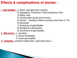
An external fistula communicating with caecum sometimes follows an operation for gangrenous appendicitis or the draining of appendix abscess. A foecal fistula can occur from necrosis of a gangrenous patch of intestine after the relief of a strangulated hernia, or from a leak from an intestinal anastomosis. The opening of an abscess connected with chronic divericulitis or carcinaoma of colon frequently results in a faecal fistula. Radiation damage can also cause fistula Most common cause is previous surgery with adhesions following previous operations. Damage to the small intestine occurs inadvertently during dissection of adhesions and because of associated sub acute obstruction or abscess.
Enterocutaneous fistulae can be divided into: 1. those with a high output, > 1 litre a day 2. those with a low output, < 1 litre a day Also anatomically: 1. Simple, with a direct communication between the gut and the skin 2. Complex, those with one or more tracts that are tortuous and sometimes associated with an intervening abscess cavity halfway along the tract
The discharge from a fistula connected with the duodenum or jejunum is bile-stained and causes severe excoriation of the skin. When the ileum or caecum is involved, the discharge is fluid faecal matter. When the distal colon is affected, it is solid or semisolid faecal matter. The site of leakage and the length of the fistula can be determined by small bowel enema and barium enema, by fistulography and, most importantly, by CT of the abdomen (show abscess).
TREATMENT Very challanging in high-output fistula. Low-output fistulae can be expected to heal spontaneously, provided there is no distal obstruction. Reasons for failure to heal: 1. epithelial continuity between the gut and the skin 2. presence of active disease e.g. carcinoma at the site of anastomosis or in the tract 3. an associated complex abscess.
The abdominal wall must be protected from erosion by the use of appliances. Nil by mouth IV nutrition Fistula output chart The higher the fistula, the more skin excoriation worst in duodenal fistula. High-output fistula causes rapid dehydration and hypoproteinaemia. Vigorous fluid replacement and nutritional support needed. Drainage if intra-abdominal abscess life saving. In complex fistula bring out a defunctioning stoma upstream of the the fistula site, even if this results in a high-output stoma.
OPERATIVE TREATMENT Operative repair should be attempted only after a trial of conservative management. The surgery can be extremely technically demanding. An anastomosis should not be fashioned in the presence of continuing intra-abdominal sepsis or when the patient hypoproteinaemic.


")