Fungal Infections of Central Nervous System: Clinical Features and Etiology
Fungal infections of the central nervous system present diagnostic challenges and are considered medical emergencies. Learn about the main fungi causing CNS infections, clinical syndromes like meningitis and brain abscess, risk factors, and how fungi reach the CNS. Etiology includes a variety of fungal agents such as Candida, Aspergillus, Cryptococcus, and more. Timely diagnosis and treatment are essential to prevent severe neurological damage.

Uploaded on Mar 04, 2025 | 0 Views
Download Presentation

Please find below an Image/Link to download the presentation.
The content on the website is provided AS IS for your information and personal use only. It may not be sold, licensed, or shared on other websites without obtaining consent from the author.If you encounter any issues during the download, it is possible that the publisher has removed the file from their server.
You are allowed to download the files provided on this website for personal or commercial use, subject to the condition that they are used lawfully. All files are the property of their respective owners.
The content on the website is provided AS IS for your information and personal use only. It may not be sold, licensed, or shared on other websites without obtaining consent from the author.
E N D
Presentation Transcript
LECTURE: Fungal Infections of Central Nervous System Editing File Important Doctor s notes Extra explanation Only F or only M " " .
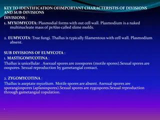
OBJECTIVES: 1. To know the main fungi that affect the central nervous system and the clinical settings of such infections. 2. To acquire the basic knowledge about fungal meningitis and brain abscess: clinical features, etiology, diagnosis, and treatment. Based on the doctor notes + foundation lectures : : Based on the doctor notes + foundation lectures FUNGI Yeast : Moulds or filamentous: Dimorphic: It s can be : septate or non-septate hypha It present in the environment inhalation It s a normal flora present in our mouths, GIT Have two forms depending on change in the environmental factors Yeast : Parasitic form, Tissue form, Cultured at 37 C Filamentous : Saprophytic form, Cultured at 25 C ( 1-Moniliaceous molds :colorless 2- dematiaceous molds : dark ) Types of fungal infections: * Superficial mycoses Cutaneous mycosis Subcutaneous mycoses Primary Systemic mycoses Opportunistic mycoses
Fungal Fungalinfections of central nervous system (CNS) : infections of central nervous system (CNS) : o CNS infections are both diagnostic challenge and medical emergency o Delay in diagnosis and initiation of appropriate therapy will lead to high mortality rate or in permanent, severe neurological damage o Fungal infections of the CNS are not common However, they are being increasingly diagnosed Why? Because of the increase of the number of immunocompromised patients. Risk Risk factors: factors: HIV/AIDS Hereditary immune defects Diabetes mellitus Immunosuppressive medications Solid organs transplantation Hematopoietic stem cell transplant (HSCT) Malignancies Neutropenia Indwelling catheters (e.g. candidemia CNS seeding) Surgery or trauma Normal flora = Opportunistic Dimorphic = Primary
How fungi reach the central nervous system: How fungi reach the central nervous system: Fungi reach the central nervous system by different mechanisms: o Hematogenous spread o Local extension from the paranasal sinuses, the ear, or the orbits. o Traumatic introduction: Injections Surgical procedures eg. Baby shunt lumbar punctures we use it to get CSF Head trauma Clinical syndromes Clinical syndromes o These clinical syndromes can occur either alone or in combination: 1- Meningitis: 2- Brain abscess: Sub acute OR Chronic With or without vascular invasion Mold Brain abscess o Certain clinical syndromes are specific for certain fungi can give us a clue Yeast Meningitis
Etiology: Etiology: Several fungal agents can cause CNS infections: Yeast: Mould: Dimorphic: Candida spp Aspergillus spp Histoplasma spp Cryptococcus spp Zygomycetes Blastomyces spp Exophiala spp Coccidioides spp Cladophialophora bantiana Paracoccidioides spp Rhinocladiella mackinziei and Others
Cryptococcal meningitis: Cryptococcal meningitis: AIDS is the leading predisposing factor * Etiology Cryptococcus neoformans is the most common etiology Capsulated yeast cells (capsulated) Naturally in Pigeon habitats (soil rich with pigeon dropping) Acquired by: Inhalation (not from human to humaun) Clinical syndromes: Mainly chronic meningitis Candidiasis: Candidiasis: normal flora Candida species are the fourth most common cause of hospital acquired blood stream infections Etiology: Candida albicans, and other species including C. glabrata, C. tropicalis C. parapsilosis, and C. krusei. Hematogenously Surgery, Catheters, trauma Cerebral abscesses Meningitis ( premature babies) can reach the CNS: Clinical syndromes: Pigeon = *Cellular immunity is important to be infected by this organism if there is any defect
CNS Aspergillosis: CNS Aspergillosis: Mortality rate is high Etiology: Aspergillus fumigatus most cause, but also A. flavus, and A. terrus Clinical syndrome: Usually brain abscesses (single or multiple) rarely as meningitis Spread Hematogenously May also occur via direct spread from the anatomically adjacent sinuses, Malignancies Transplantation Chemotherapy can reach the CNS: Common risk factors include : -it is inhaled through the respiratory system then goes to CNS through circulation or through paranasal sinuses then to CNS as local extension -Mostly effect immunocompromised pt. as chemo therapy , transplant surgery , malignancy ( opportunistic infection ) \ -very rare for immune competent
CNS CNS Zygomycosis Zygomycosis ( (mucoromycosis mucoromycosis) ) : : The rhinocerebral )it is Mucorales) form is the most frequent presenting clinical syndrome in CNS zygomycosis. Mortality is high (80- 100%) Progression is rapid, Etiology: Zygomycetes e.g. Rhizopus, Absidia, Mucor Fast growing fungi ( all of them are Mucorales ) risk factors : Diabetics with ketoacidosis because it can thrive in high acidic condition, in addition to other risk factors The clinical manifestations of the rhinocerebral form start as sinusitis, rapidly progress and involve the orbit, eye and optic nerve and extend to the brain could be inhaled Facial edema, pain, necrosis, loss of vision, black discharge Angiotropism due to blood vessel invasion; As angio-invasion is very frequent Rapid diagnosis Control the underlying disease Early surgical debridement Appropriate antifungal therapy To improve the outcome: -General term of opportunistic fungal infection caused by a class called : zygomycetes ( has two order 1-Mucorales 2-Entomophthorales.) -like Aspergillosis in the mode of transmission , start in the respiratory system then through heamatogenous to CNS -rhinocerebral has the tendency to invase blood vessel (important) usually the area involved has thrombosis , necrosis , infarction lastly will become plaque scar
Pheohyphomycosis Pheohyphomycosis: : oFungal infections caused by dematiaceous fungi darkly colored due to melanin pigment Neurotropic fungi oCNS infections: Usually brain abscess, and chronic oReported in immunocompetent hosts not immunocompromised o Etiology: Rhinocladiella mackenziei ( Mainly reported from Middle East) Cladophialophora, Exophiala , many others Other Infections: Other Infections: Histoplasmosis by Histoplasma capsulatum Blastomycosis: Blastomyces dermatitidis Coccidiodomycosis: Coccidioides immitis Paracoccidiodomycosis: Paracoccidioides brasiliensis o Histoplasmosis / Blastomycosis / Coccidiodomycosis / Paracoccidiodomycosis oCaused by primary pathogens oSub acute or chronic Meningitis (common), and brain abscess oFollowing a primary infection, mainly respiratory mainly by inhalation then through blood goes to skin , bone , CNS -Primary systemic infection = affecting the normal person / -Neurotropic = likely invading CNS / -immunocompetent = normal person
Diagnosis: Diagnosis: Clinical features (history, risk factors, etc): Neuro-imaging: radiological finding Not Specific (Past surgery is important in history ) Good value in diagnosis and therapy monitoring CSF examination (cell count, chemistry) Histopathology Lab Investigations: Microbiology Clinical Samples: CSF - Biopsy- Pus, aspirate - Blood (for serology) Lab Diagnosis: Lab Diagnosis: 1. CSF abnormalities: Not specific for Fungal infections* Cell count Glucose level (low) because it is consumed by the organism Protein level (high) We check color too (bloody , turbid) 4. PCR: the most accurate test 5. Serology: for detection of antigen or antibody Candida Aspergillus Cryptococcus Histoplasma Blastomyces Coccidioides Paracoccidioides 2. Direct Microscopy: Fungal stains: Giemsa, GMS, PAS, India ink (mostly for Cryptococcus neoformans for the presence of polysaccharide capsule ) 3. Culture: Fungal media: SDA**, BHI***, other media if needed. *because pyogenic bacteria can be the same findings in CSF ***BHI (brain & heart infusion agar **SDA (Sabouraud dextrose agar)
Lab. Diagnosis: Lab. Diagnosis: CNS infection Direct microscopy Culture Serology* Cryptococcal meningitis Yeast cells Capsulated (india ink) Yeast Cryptococcal Ag (capsule) Latex agglutination A A Candidiasis Yeast cells and pseudohyphae Yeast Manann Ag (cell wall) B B Aspergillosis Septate branching hyphae C Hyaline mould Galactomannan Ag Zygomycosis Broad non-septate hyphae D Hyaline mould Fast growing No serology available C Pheohyphomycosis Brown septate hyphae Dematiaceous mould -D- Glucan *Serology: -D- Glucan For diagnosis of invasive fungal infections except cryptococcosis and zygomycosis D Ag = antigen
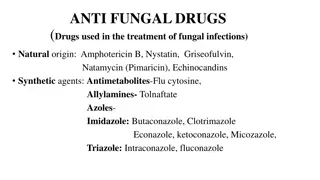
Management: Management: 1. Control of the underlying disease 2. Reduce immunosuppresion, restore immunity if possible 3. Start antifungal therapy promptly: Polyenes/ Azoles/ Echinocandins 4. Consider surgery in certain situations 5. Key of treatment is early diagnose Antifungal therapy: Antifungal therapy: CNS fungal infection Treatment Cryptoccocal meningitis Amphotericin B (combination with Flucytosine) CNS Candidiasis (some are resistance to azole ) Caspofungin, Fluconazole, Voriconazole, Amphotericin B CNS Aspergillosis Voriconazole CNS Zygomycosis Amphotericin B -Amphotericin B has side effect : renal toxicity -The Target of anti fungal : cell wall , cell membrane or DNA
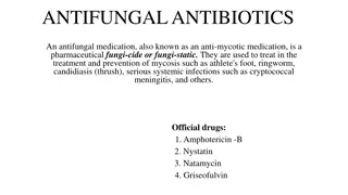
Mnemonics : ANTIFUNGAL MNEMONICS: 1- in all the antifungal we use Amphotericin B EXEPT Aspergillosis we use Voriconazole SO: Aspergillosis - Voriconazole = 2- Candidiasis - Caspofungin = LAB DIAGNOS MNEMONICS: 1- Aspergillosis - Septate branching hyphae = CNS ZYGOMYCOSIS (MUCOROMYCOSIS): 1-Etiology: Zygomycetes e.g. Absidia, Rhizopus, Mucor = +
SUMMARY: Etiology -Yeast : Candida spp, Cryptococcus spp. -Mould : Aspergillus spp, Zygomycetes. -Dimorphic : Histoplasma spp, Blastomyces spp, Coccidioides spp, Paracoccidioides spp. - AIDS ( HIV ) is the leading predisposing factor . - Acquired by inhalation. - Meningitis . - Etiology: Cryptococcus neoformans found naturally in pigeon habitats capsulated yeast cells . - Lab diagnosis : Microscope ( india ink) Capsulated . Serology Cryptococcal Ag , Latex agglutination . culture yeast. - Treatment : Amphotericin B + Flucytosine. Cryptococcal meningitis - Reach CNS by: Hematogenous spread, surgery , catheters. - Cerebral abscess and meningitis . - Etiology is mainly C.albicans(normal flora). - Lab diagnosis : Microscope psuedohyphae . Serology : Manann Ag and -D- Glucan (if invasive). Culture: yeast - Treatment : Caspofungin, Fluconazole, Voriconazole, Amphotericin B. Candidiasis - Brain abscesses . - Risk factors: Malignancy, transplantation, chemotherapy. Spread Hematogenously, or direct spread from adjacent sinuses. - Mortality rate is high . - Etiology: Aspergillus fumigatus, A.flavus and A.terrus - Lab diagnosis : Microscope Septate branching hyphae. Culture Hyaline mould. Serology Galactomannan Ag , -D- Glucan (if invasive). - Treatment : Voriconazole. CNS Aspergillosis - -The rhinocerebral form is the most frequent presenting clinical syndrome in CNS zygomycosis. - Risk factor: Diabetic with ketoacidosis . - Clinical manifestations: Start as Sinusitis rapidly progress involve the orbit eye optic nerve brain. - Symptoms: Facial edema , necrosis, loss of vision , black discharge, angiotropism . - Mortality rate is high . - etiology: Zygomycetes e.g. Rhizopus, Absidia. -Lab diagnosis : Microscope Broad non-septate hyphae. Culture Hyaline mould, fast growing . No serology test is available. - Treatment : Amphotericin B. CNS Zygomycosis ( Mucoromycosis ) -Affects immunecompetent hosts . -Caused by dematiaceous fungi (neurotropic fungi) - Chronic brain abscess . Etiology: Rhinocladiella mackenziei most common in middle east. - Lab diagnosis : Microscope Brown septate hyphae. Culture Dematiaceoud mould. Serology -D- Glucan . Pheohyphomycosis - -Etiology primary pathogens - Sub-acute or chronic meningitis (common) and brain abscess . - following a primary respiratory infection. Other infections (Histoplasmosis, Blastomycosis, Coccidiodomycosis, Paracoccidiodomycosis)
SUMMARY: - Fungal infections of CNS are not common. Associated with Meningitis (sub-acute or chronic) or brain abscess . - Risk factors: HIV , hematopoietic stem cell transplant, DM, Indwelling catheters, Solid organs transplantation, Malignancies, Neutropenia, Hereditary immune defects, Immunosuppressive medications, Surgery or trauma. -Mechanisms to reach CNS : Hematogenous spread, Traumatic introduction (e.g. lumbar punctures), Local extension from the paranasal sinuses, the ear, or the orbits. Diagnosis: Clinical features (history, risk factors, etc),Neuro-imaging, Lab Investigations Lab Investigations ( CSF examination (cell count, chemistry), Histopathology, Microbiology) CSF abnormalities Direct microscopy Culture Serology PCR Not specific for fungal infection. Fungal stains: Giemsa, GMS, PAS, India ink (Cryptococcus neoformans) Fungal media: SDA, BHI To detect the antigens
QUIZ: 1/A 54-year-old man developed a slowly worsening headache followed by gradual, progressive weakness in his right arm. A brain scan revealed a cerebral lesion. At surgery an abscess surrounded by granulomatous material was found. Sections of the tissue and subsequent culture showed darkly pigmented septate hyphae indicating phaeohyphomycosis. This infection may be caused by which species below? (A) Aspergillus (B) Cladophialophora (C) Coccidioides (D) Malassezia Answer: B 4/A patient was diagnosed with chronic brain abscess. The culturing of the organism showed Dematiceoud mould. What is the most likely causing organism? (A) Rhinocladiella Mackenzie (B) cryptococcus neoformans (C) C.albicans (D) Aspergillus fumigatus Answer: A 5/ A 52 years old male presented to the hospital with cryptococcal meningitis, history was taking and it showed that he had HIV. How did the fungus reach the CNS? (A) hematogenously (B) by inhalation (C) infection from the ear (D) infection from the paranasal sinuses Answer: B 2/A patient was diagnosed with cerebral abscess. the serology test showed mannan AG. What are we going to see under the microscope? (A)pseudohyphae (B) non- septate hyphae (C) septate hyphae (D) brown septate hyphae Answer: A 3/What is the best antifungal treatment for a patient with cytococcal meningitis? (A) Amphotercin (B) comination of amphotercin and flucytocin (C) flucytocin (D)comination of amphotercin and voriconazole Answer: B
THANK YOU FOR CHECKING OUR WORK, BEST OF LUCK! Hamad Alkhudhairy Shrooq Alsomali Shoag Alahmari Alaa Alaqeel Doctors slides