GINGIVAL SURGICAL TECHNIQUES
Dr. Sonika Bodhi presents on Gingivectomy and Gingivoplasty at Rungta College of Dental Sciences and Research, Bhilai. Explore the healing process and post-operative care involved after surgical gingivectomy. Learn about epithelial cell migration, tissue regeneration, and recovery timeline in this informative lecture.

Download Presentation

Please find below an Image/Link to download the presentation.
The content on the website is provided AS IS for your information and personal use only. It may not be sold, licensed, or shared on other websites without obtaining consent from the author. If you encounter any issues during the download, it is possible that the publisher has removed the file from their server.
You are allowed to download the files provided on this website for personal or commercial use, subject to the condition that they are used lawfully. All files are the property of their respective owners.
The content on the website is provided AS IS for your information and personal use only. It may not be sold, licensed, or shared on other websites without obtaining consent from the author.
E N D
Presentation Transcript
RUNGTA COLLEGE OF DENTAL SCIENCES AND RESEARCH,BHILAI GINGIVAL SURGICAL TECHNIQUES PRESENTED BY: Dr Sonika Bodhi Reader Dept. of Periodontology
SPECIFIC LEARNING OBJECTIVES CORE AREAS DOMAIN Cognitive CATEGORY Must to know Gingivectomy Cognitive Must to know GIngivoplasty
CONTENTS PART II Gingivectomy Gingivoplasty Summary References
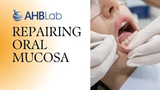
Healing after Surgical Gingivectomy The initial response ......formation of a protective surface clot; the underlying tissue becomes acutely inflamed, with some necrosis. The clot is then replaced by granulation tissue. By 24 hours, there is an increase in new connective tissue cells, mainly angioblasts, just beneath the surface layer of inflammation and necrosis.
by the third day, numerous young fibroblasts are located in the area. The highly vascular granulation tissue grows coronally, creating a new, free gingival margin and sulcus. Capillaries derived from blood vessels of the periodontal ligament migrate into the granulation tissue, and within 2 weeks, they connect with gingival vessels.
After 12 to 24 hours, epithelial cells at the margins of the wound start to migrate over the granulation tissue, separating it from the contaminated surface layer of the clot. In 24 to 36 hours....Epithelial activity at the margins reaches a peak
The new epithelial cells arise from the basal and deeper spinous layers of the wound edge epithelium and migrate over the wound over a fibrin layer that is later resorbed and replaced by a connective tissue bed. The epithelial cells advance by a tumbling action, with the cells becoming fixed to the substrate by hemidesmosomes and a new basement lamina.
After 5 to 14 days, surface epithelialization is generally complete. During the first 4 weeks after gingivectomy, keratinization is less than it was before surgery. Vasodilation and vascularity begin to decrease after the fourth day of healing and appear to be almost normal by the sixteenth day.
Complete epithelial repair takes about 1 month. Complete repair of the connective tissue takes about 7 weeks.
The flow of gingival fluid in humans is initially increased after gingivectomy and diminishes as healing progresses.
Gingivectomy by Electrosurgery Advantages Electrosurgery permits an adequate contouring of the tissue and controls hemorrhage
Disadvantages cannot be used in patients who have noncompatible or poorly shielded cardiac pacemakers. The treatment causes an unpleasant odor. If the electrosurgery point touches the bone, irreparable damage can be done
Indications limited to superficial procedures such as removal of gingival enlargements, gingivoplasty, relocation of frenum and muscle attachments, and incision of periodontal abscesses and pericoronal flaps; extreme care should be exercised to avoid contacting the tooth surface.
TECHNIQUE The removal of gingival enlargements and gingivoplasty is performed with the needle electrode, supplemented by the small, ovoid loop or the diamond-shaped electrodes for festooning. A blended cutting and coagulating (fully rectified) current is used. In all reshaping procedures, the electrode is activated and moved in a concise shaving motion
In the treatment of acute periodontal abscesses, the incision to establish drainage can be made with the needle electrode without exerting painful pressure. The incision remains open because the edges are sealed by the current. After the acute symptoms subside, the regular procedure for the treatment of the periodontal abscess is followed.
For hemostasis, the ball electrode is used. Hemorrhage must be controlled by direct pressure (using air, compress, or hemostat) first; then the surface is lightly touched with a coagulating current. Electrosurgery is helpful for the control of isolated bleeding points. Bleeding areas located interproximally are reached with a thin, bar-shaped electrode
For frenum and muscle attachments relocation ...loop electrode is used. For acute pericoronitis, drainage may be obtained by incising the flap with a bent-needle electrode. A loop electrode is used to remove the flap after the acute symptoms subside
Healing after scalpel surgery & electrosurgery Some investigators reported no significant differences. Other researchers find..... delayed healing, greater reduction in gingival height, and more bone injury after electrosurgery.
LASER GINGIVECTOMY Most often used : Carbon dioxide (CO2) ....10,600 nm neodymium:yttrium-aluminum-garnet (Nd:YAG)...1064 nm
Gingivectomy by Chemosurgery Chemicals such as 5% paraformaldehyde or potassium hydroxide not currently used.
Disadvantages: The depth of action cannot be controlled, and therefore healthy attached tissue underlying the pocket may be injured. Gingival remodeling cannot be accomplished effectively. Epithelialization and re-formation of the junctional epithelium and reestablishment of the alveolar crest fiber system occur more slowly in chemically treated gingival wounds than in those produced by a scalpel.
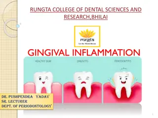
GINGIVOPLASTY Gingivoplasty is a reshaping of the gingiva to create physiologic gingival contours, with the sole purpose of recontouring the gingiva in the absence of pockets. Gingivectomy is performed to eliminate periodontal pockets and includes reshaping as part of the technique.
Gingival and periodontal disease often produce deformities in the gingiva that interfere with normal food excursion, collect plaque and food debris, and prolong and aggravate the disease process. Such deformities include (1) gingival clefts and craters, (2) shelflike interdental papillae caused by acute necrotizing ulcerative gingivitis, and (3) gingival enlargements
Gingivoplasty may be done with a periodontal knife, a scalpel, rotary coarse diamond stones, or electrodes.
It consists of procedures that resemble those performed in festooning artificial dentures: tapering the gingival margin, creating a scalloped marginal outline, thinning the attached gingiva, and creating vertical interdental grooves and shaping the interdental papillae to provide sluiceways for the passage of food
SUMMARY Current periodontal surgery must consider the 1. conservation of keratinized gingiva, 2. minimal gingival tissue loss to maintain esthetics 3. adequate access to the osseous defects for definitive defect correction, and 4. minimal postsurgical discomfort and bleeding by attempting surgical procedures that will allow primary closure
The gingivectomy surgical technique has limited use in current surgical therapy because it does not satisfy these considerations in periodontal therapy. The clinician must carefully evaluate each case as to the proper application of this surgical procedure used in different ways.
REFERENCES Newman MG, Takei HH, Klokkevold PR, Carranza FA. Carranza s clinical periodontology, 10th ed. Saunders Elsevier; 2007. Lindhe J, Lang NP and Karring T. Clinical Periodontology and Implant Dentistry. 6th ed. Oxford (UK): Blackwell Publishing Ltd.; 2015. Newman MG, Takei HH, Klokkevold PR, Carranza FA. Carranza s clinical periodontology, 13th ed. Saunders Elsevier; 2018.