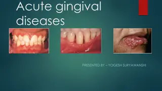
GINGIVAL INFLAMMATION

Inflammation of the gingiva, known as gingivitis, is a common precursor to periodontal diseases. This article delves into the stages of gingivitis, from initial vascular changes to advanced lesions. Explore the specific learning objectives and clinical manifestations associated with each stage, as well as microscopic signs of acute inflammation in the gingival tissue. Gain insights into the importance of early detection and treatment of gingivitis to prevent progression to more severe periodontal conditions.

Download Presentation

Please find below an Image/Link to download the presentation.
The content on the website is provided AS IS for your information and personal use only. It may not be sold, licensed, or shared on other websites without obtaining consent from the author.If you encounter any issues during the download, it is possible that the publisher has removed the file from their server.
You are allowed to download the files provided on this website for personal or commercial use, subject to the condition that they are used lawfully. All files are the property of their respective owners.
The content on the website is provided AS IS for your information and personal use only. It may not be sold, licensed, or shared on other websites without obtaining consent from the author.
E N D
Presentation Transcript
RUNGTA COLLEGE OF DENTAL SCIENCES AND RESEARCH,BHILAI GINGIVAL INFLAMMATION DR. PUSHPENDRA YADAV sr. lecturer DEPT. OF PERIODONTOLOGY 1
SPECIFIC LEARNING OBJECTIVES CORE AREAS Introduction Classification Stages of Gingivitis DOMAIN Affective Cognitive Cognitive CATEGORY Desire to know Must to know Must to know Cognitive Must to know Stage I: The Initial Lesion Stage II: The Early Lesion Cognitive Must to know Stage III: The Established Lesion Stage IV: Advanced Lesion 2
content content Introduction Classification Stages of Gingivitis Stage I: The Initial Lesion Stage II: The Early Lesion Stage III: The Established Lesion Stage IV: Advanced Lesion 3
Introduction Introduction Inflammation of gingiva is called gingivitis. Most forms of periodontal diseases are plaque- associated disorders which start as an overt inflammation of the gingiva. In susceptible individuals, if inflammation is left untreated it may spread to involve deeper portions of the periodontium. 4
STAGE I GINGIVITIS: THE INITIAL LESION vascular changes dilated capillaries and increased blood flow. occur in response to microbial activation of resident leukocytes and the subsequent stimulation of endothelialcells. Clinically, this initial response of the gingiva to bacterial plaque (subclinical gingivitis) is not apparent 7
Microscopically Signs of acute inflammation seen in the connective tissue beneath the junctional epithelium. Changes in blood vessel morphologic features (e.g., widening of small capillaries or venules) Adherence of neutrophils to vessel walls (margination) occur within 1 week and sometimes as early as 2 days after plaque accumulation. Leukocytes, mainly PMNs, leave the capillaries by migrating through the walls (diapedesis, emigration) 8
STAGE II GINGIVITIS: THE EARLY LESION The early lesion evolves from the initial lesion within about 1 week after the beginning of plaque accumulation. Clinically, no clear-cut dividing line. As time goes on, clinical signs of erythema may appear, proliferation of capillaries and increased formation of capillary loops between rete pegs or ridges 10
Bleeding on probing Between 6 and 12 days .... Increased Gingival fluid flow and transmigrating leukocytes 11
Microscopically Gingiva reveals leukocyte infiltration in the connective tissue beneath the junctional epithelium, consisting mainly of lymphocytes (75%, with the majority T cells), but also composed of some migrating neutrophils, as well as macrophages, plasma cells, and mast cells. 12
All the changes seen in the initial lesion continue to intensify with the early lesion. The junctional epithelium: densely infiltrated with neutrophils, development of rete pegs or ridges. 13
70% collagen is destroyed around the cellular infiltrate. The main fiber groups affected circular and dentogingival fiber assemblies 14
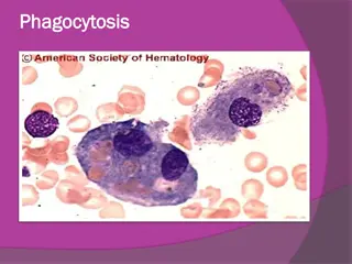
Polymorphonuclear leukocytes (PMNs) leaves the blood vessels cross the basement lamina, found in the epithelium, emerging in the pocket area. PMNs engulf bacteria in the process of phagocytosis. Fibroblasts show cytotoxic alterations, with a decreased capacity for collagen production 15
STAGE III GINGIVITIS: THE ESTABLISHED LESION predominance of plasma cells and B lymphocytes (IgG1 and IgG3) subclasses and probably in conjunction with the creation of a small gingival pocket lined with a pocket epithelium. 17
In chronic gingivitis, which occurs 2 to 3 weeks after the beginning of plaque accumulation, the blood vessels become engorged and congested, venous return is impaired, and the blood flow becomes sluggish. The result is localized gingival anoxemia, which superimposes a somewhat bluish hue on the reddened gingiva 18
Extravasation of erythrocytes into the connective tissue and breakdown of hemoglobin into its component pigments can also deepen the color of the chronically inflamed gingiva. described as moderately to severely inflamed gingiva 19
Histologically increased number of plasma cells- preponderant inflammatory cell type. Plasma cells invade deeply into the connective tissue, around blood vessels, and between bundles of collagen fibers 20
The junctional epithelium reveals widened intercellular spaces filled with granular cellular debris, including lysosomes derived from disrupted neutrophils, lymphocytes, and monocytes. The lysosomes contain acid hydrolases that can destroy tissue components. 21
The junctional epithelium develops rete pegs or ridges that protrude into the connective tissue, and the basal lamina is destroyed in some areas. In the connective tissue, collagen fibers are destroyed around the infiltrate of intact and disrupted plasma cells, neutrophils, lymphocytes, monocytes, and mast cells 22
An inverse relationship appears to exist between the number of intact collagen bundles and the number of inflammatory cells. Collagenolytic activity is increased in inflamed gingival tissue by the enzyme collagenase. Collagenase is normally present in gingival tissues and is produced by some oral bacteria and by PMNs 23
Enzyme histochemistry studies have shown that chronically inflamed gingivae have elevated levels of acid and alkaline phosphatase, -glucuronidase, - glucosidase, -galactosidase, aminopeptidase and cytochrome oxidase. esterases, Neutral mucopolysaccharide levels are decreased, presumably as a result of degradation of the ground substance 24
Established lesions of two types and appear to be reversible; some remain stable and do not progress for months or years, and others seem to become more active and to convert to progressively destructive lesions. 25
STAGE IV GINGIVITIS: THE ADVANCED LESION Extension of the lesion into alveolar bone characterizes a fourth stage known as the advanced lesion or phase of periodontal breakdown 27
Microscopically there is fibrosis of the gingiva widespread manifestations of inflammatory and immunopathologic tissue damage. plasma cells continue to dominate the connective tissues, and neutrophils continue to dominate the junctional epithelium and gingival crevice 28
summary Gingival conditions/diseases that mainly involve interdental papilla and gingival margin are gingival abscess, necrotizing ulcerative gingivitis, linear gingival erythema and drug induced gingival enlargement. Gingival ulcers are usually seen in NUG, herpes simplex virus stomatitis, aphthae, self-injury, malignant neoplasms, drugs, dermatoses, systemic diseases (hematological disorders, tuberculosis, syphilis, herpes virus, HIV). 29
REFERENCE Newman MG, Takei HH, Klokkevold PR, Carranza FA. Carranza s clinical periodontology, 10th ed. Saunders Elsevier; 2007. Lindhe J, Lang NP and Karring T. Clinical Periodontology and Implant Dentistry. 6th ed. Oxford (UK): Blackwell Publishing Ltd.; 2015. Newman MG, Takei HH, Klokkevold PR, Carranza FA. Carranza s clinical periodontology, 13th ed. Saunders Elsevier; 2018. 30
THANK YOU THANK YOU 31


")