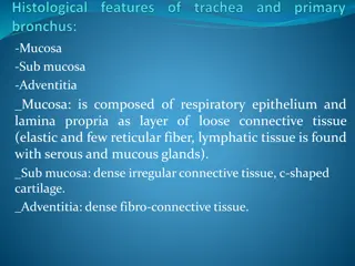
Histology of Upper Respiratory Tract
Exploring the microscopic structures of the upper respiratory tract including the nasal cavity, paranasal sinuses, and larynx. Learn about the vestibule of the nasal cavity, respiratory mucosa, nasal septum, olfactory mucosa, and more. Dive into the conducting and respiratory portions of the respiratory system. Understand the cellular composition and vascularization of the nasal cavity mucosa, along with clinical applications such as sinusitis.

Download Presentation

Please find below an Image/Link to download the presentation.
The content on the website is provided AS IS for your information and personal use only. It may not be sold, licensed, or shared on other websites without obtaining consent from the author. If you encounter any issues during the download, it is possible that the publisher has removed the file from their server.
You are allowed to download the files provided on this website for personal or commercial use, subject to the condition that they are used lawfully. All files are the property of their respective owners.
The content on the website is provided AS IS for your information and personal use only. It may not be sold, licensed, or shared on other websites without obtaining consent from the author.
E N D
Presentation Transcript
( RESPIRATORY SYSTEM ( I Histology of the Upper Respiratory Tract (Nasal cavity, Paranasal sinuses (and Larynx
Objectives: By the end of this lecture the student should be able to describe the microscopic structures of: Vestibule of the nasal cavity. Respiratory mucosa of the nasal cavity. Nasal septum. Olfactory mucosa of the nasal cavity. Mucosa of the paranasal sinuses. Larynx.
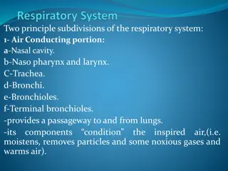
RESPIRATORY SYSTEM Conducting portion : Nasal cavity. Nasopharynx. (A 1 2 3 4 5 Larynx. Trachea. Primary bronchi (extrapulmonary bronchi). 6- Intrapulmonary bronchi: 2ry bronchi (lobar bronchi). (segmental bronchi). 7- Primary bronchioles (preterminal bronchioles). 8- Terminal bronchioles. Respiratory portion: Respiratory bronchioles. - - 3ry bronchi A) 1- 2- 3 4 Alveolar ducts . Alveolar sacs. Pulmonary alveoli.
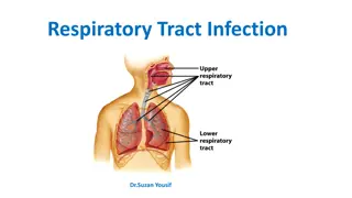
NASAL CAVITY (N.C.) Anterior portion of N.C.: .1 Vestibule. Posterior portion of N.C.: Respiratory region. Olfactory region. .2 a- b- N.B. The nasal septum divides the nasal cavity into two halves (right and left).
.VESTIBULE OF N.C Lining: is lined with thin skin. Epidermis: (Keratinized stratified Squamous epithelium). 1 2 Dermis. Contents: Vibrissae: stiff hairs. Sebaceous glands. Sweat glands. 1 2 3 Wall: 1 2 Hyaline cartilage. Cancellous (spongy) bone.
RESPIRATORY REGION (AREA) OF NASAL CAVITY MUCOSA (MUCOUS MEMBRANE): Respiratory Epithelium: Pseudo-stratified ciliated columnar epithelium with goblet (A) cells. Main Types of cells ( all touch the basement membrane) 1- Ciliated columnar cells. 2- Goblet cells. 3 4 Basal cells: are stem cells. DNES cells: e.g. serotonin.
(B) Lamina propria ( Sub-epithelial C.T.): contains: 1- Large arterial plexuses & venous sinuses (Highly vascularized C.T.) 3- Many seromucous glands (acini). Abundant lymphoid elements: Including occasional lymphoid nodules, plasma cells & mast cells. 4-
PARANASAL SINUSES Lining: 1- Respiratory epith. (Mention .) 2- Lamina propria. CLINICALAPPLICATION: Sinusitis.
OLFACTORY REGION (AREA) OF NASAL CAVITY (OLFACTORY MUCOSA) Site: 1-Roof of nasal cavity. 2-Upper part of nasal septum. 3-over superior concha. Structure: Olfactory epithelium: A) Pseudo-stratified columnar epith. 1- Olfactory cells (olfactory nerve cells) 2- Sustentacular (supporting) cells. 3- Basal cells: Pyramidal in shape, basal in position and act as stem cells.
Lamina propria: contains: Highly (richly) vascularized loose C.T. (B) 1 2- Contents: Bowman s glands ( olfactory glands) : (a are serous acini. Bundles of unmyelinated nerve fibers: Are axons of olfactory nerve (b cells + Schwann-like cells (glial cells). Rich vascular plexus. Numerous lymphoid elements. (c (d
OLFACTORY EPITHELIUM 1- Olfactory cells: Are bipolar neurons Dendrite has olfactory vesicle that has nonmotile cilia. Axons are unmyelinated with Schwann-like cells. Axons will collect in the lamina propria to form bundles of nerve fibers. Bundles will collect to form the olfactory nerve. 2- Sustentacular (supporting) cells: Are columnar cells. Function: Physical support and nourishment for olfactory cells.
LARYNX (A) Mucosa (Mucous membrane ): Epithelium. Lamina propria. 1 2 (B) Cartilages. Extrinsic and intrinsic muscles: all are skeletal. (C) Ligaments. (D)
LARYNX Mucosa: (A Epithelium: (2 types) Respiratory epithelium: 1 a- Pseudostratified ciliated columnar epithelium with goblet cells. Non keratinized stratified squamous epithelium: b- -Vocal folds. In: Superior surface of epiglottis - 2 Lamina propria.
LARYNX Mucosa (cont.): (A) There are 2 pairs of shelf-like mucosal folds: Vestibular folds: 1 Are immovable. L/M: a- epithelium. b- Lamina propria: Respiratory Loose C.T. with seromucous glands lymphoid elements & adipose cells. VOCAL FOLDS (CORDS): have: Epithelium: non keratinized stratified squamous. Lamina propria: C.T. containing bundles of elastic 2 .a .b fibers and skeletal muscle . N.B. No lymphoid nodules, No seromucous glands.
(B) Cartilages: 1- Hyaline cartilages: e.g. Thyroid cartilage. 2- Elastic cartilages: Epiglottis. (C) (D) Muscles: all are skeletal. Ligaments.


")

 OF NASAL")
 Lamina propria ( Sub-epithelial C.T.):")

 OF NASAL CAVITY")


 Cartilages:")