Intrauterine Growth Restriction (IUGR)

Intrauterine Growth Restriction (IUGR) is a condition where fetal weight falls below the 10th percentile for gestational age, impacting cellular growth processes and contributing factors such as maternal, placental, and fetal influences. This guide outlines the phases of fetal growth, etiology of IUGR, and factors affecting maternal, placental, and fetal aspects related to this condition.

Download Presentation

Please find below an Image/Link to download the presentation.
The content on the website is provided AS IS for your information and personal use only. It may not be sold, licensed, or shared on other websites without obtaining consent from the author.If you encounter any issues during the download, it is possible that the publisher has removed the file from their server.
You are allowed to download the files provided on this website for personal or commercial use, subject to the condition that they are used lawfully. All files are the property of their respective owners.
The content on the website is provided AS IS for your information and personal use only. It may not be sold, licensed, or shared on other websites without obtaining consent from the author.
E N D
Presentation Transcript
Definition: Is a fetal weight that is below the 10th percentile for gestational age as determined through an ultrasound. This can also be called small-for gestational age (SGA) or fetal growth restriction.
Phases of fetal growth First 16 weeks: mostly cellular hyperplasia 16-32 weeks: both hyperplasia and hypertrophy >32 weeks: mostly hypertrophy Thus: early growth restriction will affect cell numbers and have a global (symmetrical IUGR) effect. Later cell size will be affected (asymmetrical IUGR)
Etiology of IUGR Maternal Factors Placental Factors Fetal Factors
Maternal Factors 1. Genetic size 2. Demographics: Age (extremes of reproductive age) Socioeconomic status 3. Underweight before pregnancy or malnutrition 4. Exposure to teratogens (drugs, radiation, etc.)
5. Factors that interfere with placental flow and function: -Heart disease -Renal disease -Hypertension -Pulmonary disease -Hemoglobinopathies -Collagen-vascular disease -Diabetes -Postmaturity
-Multiple gestation -Uterine anomalies -Thrombotic disease -High altitude environment -Smoking -Cocaine
Placental Factors -Malformations -Abruption -Previa -Abnormal trophoblast invasion
Fetal Factors -Constitutional genetically small, but genetically normal -Chromosomal abnormality only about 5% of SGA babies -Malformations CNS, skeletal -Congenital infections CMV, rubella
Characteristics of IUGR symmetric: 1. Early onset 2. Head circumference, length &weight are all proportionally reduced for gestational age(below 10thpercentile) 3. Could be constitutional or normal small. 4. Examples: -Genetic causes, chromosomal -TORCH infections
Asymmetrical : 1. Late onset 2nd-3rdtrimester. 2. Growth arrest. 3.Brain sparing ,which means transfer of oxygen & nutrients to the brain .This allows normal brain and head growth while diminished glucose transfer and hepatic storage would primarily affect cell size and not number, and fetal abdominal circumference which reflects liver size would be reduced.
4. Examples: -Chronic hypoxia -Preeclampsia, chronic hypertension -malnutrition
Diagnosis of IUGR Abdominal examination: Serial Fundal height measurement in centimeters usually corresponds with the number of weeks of pregnancy after the 20th week. If the measurement is low for the number of weeks (2-3 cm), the baby may be smaller than expected.
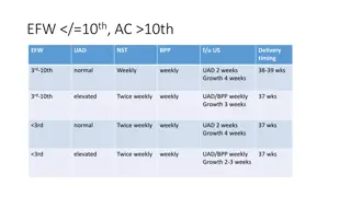
Ultrasound more accurate method of estimating fetal size. Measurements can be taken of the fetus' head and abdomen and compared with a growth chart to estimate fetal weight. The fetal abdominal circumference is a helpful indicator of fetal nutrition. , obtaining a second growth assessment over a 2- to 4-week interval is important unless strong supportive data or risk factors warrant an immediate change in management plans.
Amniotic fluid volume An association between pathological fetal-growth restriction and oligohydramnios has long been recognized. Hypoxia and diminished renal blood flow explain oligohydramnios
Doppler flow Doppler velocimetry is considered standard in the evaluation and management of the growth-restricted fetus. abnormal umbilical artery Doppler velocimetry findings characterized by absent or reversed end-diastolic flow have been uniquely linked with fetal-growth restriction. Other modality of Doppler velocimetry, include middle cerebral arteries & ductus venosus assessment
Important points regarding managements: 1.If fetal-growth restriction is suspected, then efforts are made to confirm the diagnosis, assess fetal condition, and search for possible causes. 2. antepartum fetal surveillance should include Daily fetal heart rate tracings, weekly Doppler velocimetry, and sonographic assessment of fetal growth every 3 to 4 weeks are initiated
3. growth-restricted fetuses may not tolerate the metabolic effects of corticosteroids in the same way as an unstressed fetus, increased surveillance during administration. 4. timing of delivery is crucial, and the risks of fetal death versus the hazards of preterm delivery must be considered. 5. delivery between 34 and 37 weeks when there are concurrent conditions such as oligohydramnios. With a reassuring fetal heart rate pattern, vaginal delivery is planned
However, some of these fetuses do not tolerate labor, necessitating cesarean delivery,why? Answer: - Fetal-growth restriction is commonly the result of placental insufficiency , this condition is likely aggravated by labor - diminished amnionic fluid volume increases the likelihood of cord compression during labor
Early neonatal morbidities in IUGR Birth asphyxia Meconium aspiration Hypoglycemia Hypocalcemia Hypothermia Polycythemia,hyperbilirubiemia
Thrombocytopenia Pulmonary haemorrhage Malformation Sepsis Respiratory distress syndrome Necrotizing enterocolitis. Later in life Hypertension and atherosclerosis.


![BCH 462- Biotechnology & Genetic engineering [Practical]](/thumb/273367/bch-462-biotechnology-genetic-engineering-practical.jpg)