Laboratory Diagnosis of Upper Respiratory Tract Infections

Respiratory tract infections can affect the upper respiratory tract, causing conditions like pharyngitis, streptococcal infections, and diphtheria. Learn about the common bacterial causes, diagnostic techniques, and cellular morphology associated with these infections.

Download Presentation

Please find below an Image/Link to download the presentation.
The content on the website is provided AS IS for your information and personal use only. It may not be sold, licensed, or shared on other websites without obtaining consent from the author.If you encounter any issues during the download, it is possible that the publisher has removed the file from their server.
You are allowed to download the files provided on this website for personal or commercial use, subject to the condition that they are used lawfully. All files are the property of their respective owners.
The content on the website is provided AS IS for your information and personal use only. It may not be sold, licensed, or shared on other websites without obtaining consent from the author.
E N D
Presentation Transcript
Laboratory Diagnosis LECTURE 10
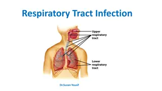
Respiratory Tract Infections Respiratory tract infections divided in to: Upper Respiratory tract infections Lower Respiratory tract infections Upper respiratory tract infections The upper respiratory tract extends from the larynx to the nostrils and comprises the oropharynx and the nasopharynx together with the communicating cavities, the sinuses and the middle ear. The upper respiratory tract can be the site of several types of infection: pharyngitis, sometimes involving tonsillitis, and giving rise to a sore throat nasopharyngitis otitis media sinusitis epiglottitis
Bacterial infections: Pharyngitis Streptococcus pyogenesis by far the most frequent cause of bacterial pharyngitis and tonsillitis. This infection is particularly prevalent in young children (5 12 years). When streptococcal pharyngitis is associated with a characteristic skin rash, the patient is said to have scarlet fever. In infants, a streptococcal throat infection may often involve the nasopharynx and be accompanied by a purulent nasal discharge.
Diphtheria Corynebacterium diphtheria is the cause of diphtheria, Diphtheria is a serious disease. C. diphtheria causes a typical form of infection, characterized by a greyish-white membrane at the site of infection (pharynx, tonsils, or larynx). Culture for Corynebacterium diphtheriae Although the diphtheria bacillus grows well on ordinary blood agar, growth is improved by inoculating one or two special media: L ffler coagulated serum or Dorset egg medium. Both of these media give abundant growth of the diphtheria bacillus after overnight incubation.
Moreover, the cellular morphology of the bacilli is more typical : irregularly stained, short to long, slightly curved rods, showing metachromatic granules, and these bacteria arranged like Chinese letters. Metachromatic granules are more apparent after staining with Albert stain by which the bacillus bacteria stained green while the metachromatic granules stained brown.
Gonococcal pharyngitis Culture of throat swabs for gonococci (Neisseria gonorrhoeae) should be done on specific request from the clinician, using the appropriate selective medium (Thayer Martin medium). Whooping Cough (Pertussis) Whooping cough is the common name for pertussis, The disease is caused by Bordetella pertussis, a tiny, encapsulated, strictly aerobic, Gram-negative rod. These organisms do not tolerate drying or sunlight and die quickly outside the host. laboratory diagnosis include culturing of nasopharyngeal swabs on a selective media (Bordet-Gengou medium).
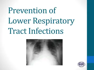
Lower respiratory tract infections Lower respiratory tract infections (LRTI) are infections occurring below the level of the larynx, i.e. in the trachea, the bronchi, or in the lung tissue (tracheitis, bronchitis, lung abscess, pneumonia). The most common infections Pneumonia Causative agent of Pneumonia is Streptococcus pneumoniae, the pneumococcus, is a Gram- positive diplococcus. The most striking characteristic of S. pneumoniae is its thick polysaccharide capsule, which is responsible for the organism s virulence. This infection is nearly always caused by S. pneumoniae. A rare cause of a rather similar form of pneumonia is Klebsiella pneumoniae. Other Gram-negative rods also can cause pneumonia, especially if host defenses are impaired.
pneumococcus, Gram- positive diplococcus
Pulmonary tuberculosis The sputum of patients with pulmonary tuberculosis( Mycobacterium tuberculosis)should be examined microscopically for acid-fast stained smear (Ziehl Neelsen) to detect immediately any patients who have acid-fast bacteria in their sputum. After the smear has been stained, the sputum should be treated by a decontamination procedure with 4% sodium hydroxide to kill contaminating bacteria and preserve mycobacteria without much effect on their survival and thus suitable for culture on L wenstein Jensen medium
Collection of sputum specimens The sputum should be collected in a sterile wide- mouthed container with a secure, tight-fitting cover and sent to the laboratory. Label the specimen with the patient's name, the date and time collected. Send the specimen to the laboratory immediately. Refrigerate the specimen if a delay of greater than one to two hours is anticipated. Microscopic examination A portion of the purulent or mucopurulentsputum should be used for the preparation of a Gram-stained smear. Gram-stained smear may provide guidance to the clinician in the choice of antimicrobial chemotherapy
Sputum Culture Sputum or phlegm is the mucousy substance secreted by cells in the lower airways (bronchi and bronchioles) of the respiratory tract. It differs from saliva, which is produced higher up, in the mouth. Sputum can be any color including clear, white, yellow, green, pink or red and blood tinged with different medical conditions. In addition to containing dead cells, foreign debris that is inhaled into the lung, and at times, bacteria, sputum contains white blood cells and other immune cells that protect the airway from infections. By using a sterile loop sputum inoculate on to the various culture plates.
A suggested routine set of culture media is as follows: Blood agar, with a streak of S. aureus to facilitate satellite growth of H. influenzae, and with an optochin disc placed in the middle of the secondary streaking, Chocolate agar. MacConkey agar. The blood agar and chocolate agar plates are incubated at 36 37 C in an atmosphere containing CO2 (e.g. in a candle jar) and the MacConkey plate is incubated in air. Sabouraud dextrose agar used for the isolation of fungi.
Gastrointestinal Tract Infections (GTI) Enteric bacterial infections, causing diarrhoea, dysentery, and enteric fevers are important health problems throughout the world. Diarrhoeal infections are second only to cardiovascular diseases as a cause of death, and they are the leading cause of childhood death. Etiological agents The etiological agents which causing gastrointestinal tract infections divided in to: Bacterial infections : The genus Salmonella cause gastroenteritis and typhoid fever Shigella spp. are the main cause of bacterial bacillary dysentery diarrhoea-producing Escherichia coli Vibrio cholerae cause Cholera, Campylobacter jejuni Clostridium difficile is the primary cause of enteric disease related to antimicrobial therapy. It produces a broad spectrum of diseases ranging from mild diarrhoea to potentially fatal pseudomembranous colitis. Viral diarrheas: Rotavirus is a major cause of diarrheal disease in children. Parasitic diarrheas: Entamoebahistolytica and Giardia lamblia can cause of diarrheal disease.
Media for enteric pathogens For Shigella spp., Salmonella spp. and Y. enterocolitica, MacConkey agar with crystal violet is recommended as a general purpose medium. Xylose lysine deoxycholate (XLD) agar is recommended for the isolation of Shigella and Salmonella. Hektoen enteric agar (HEA) or Salmonella Shigella (SS) agar are suitable alternatives. For Campylobacter spp. there are several selective media (Blaser, Butzler, Skirrow) containing different antimicrobial supplements used. Thiosulfate citrate bile salts sucrose (TCBS) agar is selective for V. cholerae. Cefoxitin cycloserine fructose agar (CCFA) is selective for Clostridium difficile
After inoculation of these media with one loopful of the faecal suspension, incubate the agar plates. Incubate the plates for the isolation of Salmonella, Shigella and Yersinia spp. and V. cholerae at 35 C in anaerobic incubator (without CO2), the plates for Campylobacter spp. at 42 C in an microaerophilic atmosphere with 10% CO2, and the plates for Clostridium difficile at 35 C in an anaerobic atmosphere


")