Microhematocrit Procedure and Reference Intervals
Microhematocrit, also known as packed cell volume (PCV), is a simple procedure to measure the ratio of red blood cells in whole blood. Learn how to perform the microhematocrit test using a centrifuge and capillary tubes. Reference intervals for different population groups are provided for interpretation.

Download Presentation

Please find below an Image/Link to download the presentation.
The content on the website is provided AS IS for your information and personal use only. It may not be sold, licensed, or shared on other websites without obtaining consent from the author.If you encounter any issues during the download, it is possible that the publisher has removed the file from their server.
You are allowed to download the files provided on this website for personal or commercial use, subject to the condition that they are used lawfully. All files are the property of their respective owners.
The content on the website is provided AS IS for your information and personal use only. It may not be sold, licensed, or shared on other websites without obtaining consent from the author.
E N D
Presentation Transcript
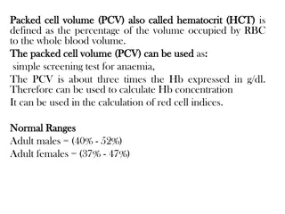
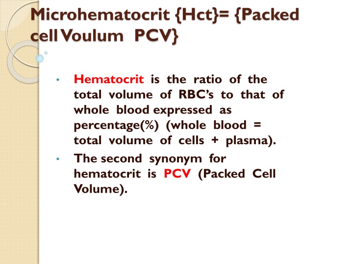
Microhematocrit {Hct}= {Packed cell Voulum PCV} Hematocrit is the ratio of the total volume of RBC s to that of whole blood expressed as percentage(%) (whole blood = total volume of cells + plasma). The second synonym for hematocrit is PCV (Packed Cell Volume).
Principle The procedure is easy to perform, whole blood is centrifuged in a narrow tube (capillary tube directly from a finger prick, to a heparin coated capillary tube.) cellular elements will be separated from the plasma, after centrifugation blood will be separated into 3 layers :
Apparatus and Materials: 1- Microhematocrit centrifuge. 2- Modeling clay (seal material). 3- Capillary tubes (7 cm long, 1mm diameter) 4- Hematocrit measuring device reader or a conventional ruler.
Procedure: 1- Fill the capillary tube with blood by capillary attraction. Either from freeflowing finger punctured by a sterile lancet/ or from a well mixed anticoagulated whole venous blood (this requires only few microliters of blood). 2- Seal with the modeling clay the empty end of the capillary tube.
Procedure: 3- Place and position the capillary tube in the radial grooves of the microhematocrit centrifuge with the sealed end away from the center (pointed toward the outside). 4- Centrifuge for 5 minutes at 10000 rpm .
Procedure: 5- The height of the RBC column, and the total column should be measured with the aid of a ruler in cm and mm, then divide the RBC column height over the total column height (total height = RBC column + buffy coat + plasma column). 6- Express the results in percentage (%).
Buffy coat is the layer where WBCs and Platelets are collected to, after centrifuging a whole blood sample, this is the middle whitish-tan colored layer.
Reference intervals: -Males : 40 - 53% Females : 37 - 47% Newborns: 51 - 60% Children : 34 - 49%
Reference intervals: Nowadays, Hct is supplied by the widely used automated hematology analyzers. But this Hct is calculated rather than measured, these analyzers are not equipped with centrifuges,
PCV RBCs =---------------- *106 6 PCV Hb =--------------- gm/100 cc 3
NOTE PCV with normal condition in pregnancy due to { body fluid in pregnant woman lead to plasma volume }. PCV in case of polycythemia and loosing fluid such as diarrhea ,barns ,vomiting. PCV in Anemia and bleeding.