Optimizing Flow Cytometry Experiments: Antibody Titration and Signal Optimization
Explore the key considerations in designing and controlling experiments, focusing on sample preparation, marker selection, and optimizing flow cytometry experiments. Learn about the impact of cell preparation methods, marker expression levels, and reagent titration on obtaining biologically meaningful results. Discover tips for achieving the best possible separation between positive and negative signals in flow cytometry analysis.

Download Presentation

Please find below an Image/Link to download the presentation.
The content on the website is provided AS IS for your information and personal use only. It may not be sold, licensed, or shared on other websites without obtaining consent from the author. If you encounter any issues during the download, it is possible that the publisher has removed the file from their server.
You are allowed to download the files provided on this website for personal or commercial use, subject to the condition that they are used lawfully. All files are the property of their respective owners.
The content on the website is provided AS IS for your information and personal use only. It may not be sold, licensed, or shared on other websites without obtaining consent from the author.
E N D
Presentation Transcript
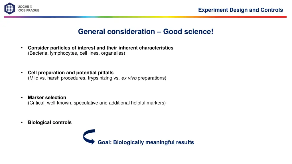
Experiment Design and Controls General consideration Good science! Consider particles of interest and their inherent characteristics (Bacteria, lymphocytes, cell lines, organelles) Cell preparation and potential pitfalls (Mild vs. harsh procedures, trypsinizing vs. ex vivo preparations) Marker selection (Critical, well-known, speculative and additional helpful markers) Biological controls Goal: Biologically meaningful results
Experiment Design and Controls Sample preparation: Cell preparation ex vivo can affect quality of samples significantly Harsh procedures can kill cells and/or damage displayed markers Harshness Scale: Trypsin > Collagenase A > Dispase > Liberase > Collagenase D Needs to be tested empirically for specific organs and markers of interest Blood cells Bacteria o Organells o Cell lines o Extracted cells from tissue (often harsh procedures required) o Whatever protocol you are using You should end up with a single cell suspension !
Experiment Design and Controls Optimizing your experiment: Flow cytometry consideration 1. Reagent optimization (titration) Background and financial considerations 2. Doublet and dead cell exclusion Measuring of live, single cells 3. Signal amplification Setting the correct PMT voltage 4. Choice of fluorochromes & spectral overlap Compensation 5. Controls in flow cytometry Which controls are needed? Goal: Best possible results
Optimizing your experiment 1. Reagent use & antibody titration Goal: best possible separation between positive and negative signals What are the fluorescent markers in my sample? Intrinsic fluorescent markers (e.g. transgenic GFP mice) Extrinsic = staining majority of flow cytometry experiments Signal intensity of extrinsic markers depends on: Marker expression level (cannot be influenced) Quality of the antibody (hard to influence) Optimal reagent concentration influence by titration
Optimizing your experiment 1. Antibody titration basics maximize signal:noise (pos/neg separation) - this may occur at less then saturated staining - this may or may not be the manufacturer s recommended titer concentration too low positive signal decreases concentration too high background increases optimal concentration = minimal antibody concentration yielding maximum signal separation Protocol: prepare the dilution series (4-8) stain your cells according to your standard protocol analyze the cells on flow cytometer export data and analyze in software of choice calculate the stain index Alternative equation for Stain Index: Stain Index: ?????? ?????? 2 ? ????? ?????? ?????? [ 84%??? ??????/0.995] ?? = ?? =
Optimizing your experiment 1. Reagent use & antibody titration Decreasing concentration decreases SI because of decreased positive staining Increasing concentration decreases SI because of increasing background 1:50 Concentration of Ab 1:100 1:200 Stain Index Optimal staining range 1:400 1:800 1:1200 1:1600 CD8 - PerCP-Cy5.5 1:50 1:100 1:200 1:400 1:800 1:1200 1:1600 Concentration of Ab Consider: clone, affinity, monoclonal vs. polyclonal, Ig-Isotype, type of Fluorochrome, concentration
Optimizing your experiment 2. Dead cell exclusion Problem: Dead cells, with compromised membrane integrity, tend to be sticky false positives exclude dead cells from analysis bind all sorts of reagents unspecifically Solution: Exclude dead cells with live-dead marker I. DNA intercalators a) Propidium iodide (PI) L/D marker working principle b) 7-Aminoactinomycin D (7-AAD) c) DAPI II. Amine-reactive dyes a) different emission types availiable e.g. Zombie Aqua/Yellow/NIR These dyes can be fixed after staining!
Optimizing your experiment 3. Doublet exclusion Goal: measuring single cells Easy exclusion by exploiting relationship between signal height, area and width of the FSC and SSC Tim Bushell
Optimizing your experiment 4. Optimizing Photon Multiplier Tube Voltages (PMTVs) Goal: observe all events on scale quantifiable data PMT voltages affect signal amplification FSC & SSC PMTVs: can be adjusted to suit your preferences/cells ensure that all relevant events are observed on the scatter plot Fluorescent parameter PMTVs: Ensure that positive and negative populations are on scale Modern cytometers offer preset values which were optimized during quality control (dim, medium & bright beads) Do not change PMTVs after recording compensation controls PMTV settings based on unstained cells only necessary for old analog cytometers (FACSCalibur) NOT a valid approach for modern digital devices!
Optimizing your experiment 4. Optimizing Photon Multiplier Tube Voltages (PMTVs) Goal: observe all events on scale quantifiable data FL-1 PMTVs FL-1 FL-1 Avoid off scale populations SSC-A FCS-A
Optimizing your experiment 5. Fluorochrome choice & Compensations Goal: brightest signals, smallest spectral overlap Appropriate choice of fluorochromes to minimize spectral overlap Where unavoidable, spectral overlap requires compensation (most fluorochromes have broad emission spectra)
Optimizing your experiment 5. Fluorochrome choice & Compensations Goal: brightest signals, smallest spectral overlap process of correcting spectral overlap is a mathematical operation called compensation calculated automatically by wizard in Diva software required: single-stained controls for each fluorochrome used in an experiment usually compensation values are listed in percent e.g. PE -10% FITC " means that 10% of FITC signal (donor) are detected in the PE detector (receiving channel)! compensation value depends on: cytometer set-up (mirrors, filters, lasers) fluorophore properties (Alexa-488 vs. FITC) voltage of a given PMT Set PMT voltages before recording single stains! Note: PMTs detects photons regardless from which fluorochrome they originate
Optimizing your experiment 6. Controls in Flow Cytometry Goal:correctly compensated/gated specific data Controls must undergo the same treatment (i.e., preparation, fixation) as all the tubes in an experiment. Prior acquisition Compensation Single-stained controls Gating controls fluorescence minus one (FMO) controls Where does negative end and positive begin? Specificity controls isotype controls Post acquisition Quality control of your acquired data Stability of flow during measurement!
Optimizing your experiment Controls: a) Specificity controls Unstained controls: - to detect the autofluorescence* or background staining - to set up PMT-voltage for FSC, SSC and FL-channels *Autofluorescence: - fluorescent signals generated by the cells themselves (from pyridine and flavin nucleotides) - present in all cells (viable and dead) - adds to fluorescence label of cells > decreases fluorescence detection limit - observed in all fluorescence channels, but decreases dramatically at longer wavelengths (>600 nm, far-red/infra-red) Solution: for cell types with high autofluorescence, a dye with a longer emission wavelength (APC, APC-Cy7) often provides excellent signal-to-noise ratio Secondary controls: - for indirect staining - secondary Ab alone to control for non-specific binding of this polyclonal Ab to dead or sticky cells
Optimizing your experiments Controls: a) Specificity controls Specificity (experimental and gating) controls e.g. transfected cells: transfected / mock transfected / wt cell line primary cells: WT / KO or activated / naive Isotype controls: not necessary for (lineage) markers with clearly separated population Ab with the same Ig isotype as the test Ab, specificity known to be irrelevant to the analyzed sample Use commercially available isotype control of unknown specificity or choose suitable antibody which recognizes an antigen that presumably is not present on the cells of interest Control for nonspecific staining of an antibody of a particular isotype conjugated to a particular fluorochrome Suitable for identifying the problem, but not to quantify it do not gate based on unspecific staining!
Optimizing your experiments Tip for not using isotype controls: You certainly do not need them for things that are clearly bimodal If you are looking for T cells and B cells in peripheral blood..
Experiment Design and Controls Controls: b) Single stained-controls Goal: high quality single-stained controls Positive and negative population must have the same autofluorescence, i.e. should be of the same kind Same fluorochromes as in your experiment must be used Especially important for tandem-dyes (Lot, manufacturer, etc.)! Single-stained control should be at least as bright or brighter than experimental staining compensation beads Compensation controls should be treated the same way as samples (fixation, incubation time, etc.)
Experiment Design and Controls Controls: b) Single stained-controls Antibody capture beads as single stain controls: saves sample more precise computation of the spillover (beads have smaller signal CV) useful for parameters that are too dim in the experimental setting i.e. cytokines, chemokines, Note: Some beads are isotype specific! (Ultracomp beads by Ebiosciences are versatile) Cells as single stained-controls: work for ALL reagents (PI, DAPI, ) the control is, ideally, as bright as the experimental sample (not necessarily true) You can combine cells and beads for single-stained controls! use a controls containing unstained & stained cells or unstained & stained beads, respectively, to provide the correct negative population for the calculation
Experiment Design and Controls Controls: c) Fluorescence Minus One (FMO) controls Control which is used to identify and gate cells in the context of data spread due to the multiple fluorochromes in a given panel An FMO control contains all the flurochromes in a panel, except for the one that is being measured Ideally add an isotype control in place of the controlled reagent (FMO + isotype) Good gating controls!
Controls in Flow Cytometry Stability of the flow: Quality control post aquisition: Check stability of fluorescence signal over time for at least one fluorescence parameter/channel per laser Flow instabilities can be caused by loading of dense samples (esp. at the start of a measurement) or partial clogs pressure fluctuations Always wait a few seconds for the flow to stabilize before recording data!
Controls in Flow Cytometry Stability of the flow: "Poor flow " indicates a major issue with fluidics and if this is observed, it is time to stop and do a quick cleaning/check of the system. Cleaning tips: WASH on High Flow Rate:5 min FACS Clean >5 min FACS Rinse> 5 min H2O Remember, filtering samples is always a good idea!
Experiment design Analytical variables to consider Data Acquisition, Analysis and Interpretation Instrument setup and Interpretation use unstained control to adjust and optimize FSC & SSC PMTVs run fully-stained sample before you record compensation controls set voltages: decrease voltages for any detectors where event are off-scale increase voltages for any detectors where low-end resolution is poor run single-stained compensation controls for each experiment and set compensation (use Diva software) run appropriate controls: gating controls (e.g., FMO), biological controls (e.g., unstimulated controls) think about the speed of analysis (high flow rate less intensity resolution)
Experiment design Analytical variables to consider Data analysis / Interpretation record appropriate number of events to ensure reliable results, think about the gating strategy visually inspect your data check gating across all samples in experiment, use back gating Avoid classification errors and false conclusions due to improper compensations and/or gating, or sample artifacts Ask for interpreting the data, experiment and instrument setup save time and labor
Some general handling tips: 1. If in doubt with anything: get your core staff. 2. You can not destroy your cytometer by inserting a sample 3. You can not destroy your cytometer unless you hit / kick or flood it. 4. Always follow SOPs (Standard Operating Procedures) if you have some. 5. Rule of thumb: 1. unplugging as well as taking in and out is ok 2. unscrewing (with a screw driver) is in most of the cases NOT ok. 3. If Rule 5.2. was unfortunately violated immediately follow handling tip number 1.
Experiment Design and Controls Take home message: Planning and quality-control pays off and saves time in the long run! Always titrate your antibodies! Use singlet gating and a dead cell marker! Before you start complex experiments: test your panels! Prepare your single-stained controls perfectly! Include FMOs for questionable signals and consider isoptype controls esp. when working with new cells! Quality-control your data after acquisition (time parameter)!