Orbital Tumors and Clinical Evaluation
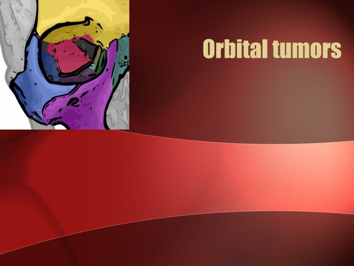
Orbital tumors can present with proptosis, pain, and vision loss, necessitating surgical intervention in cases affecting vision or optic nerve compression. Complications may include ptosis, diplopia, and visual loss. Differentiate classifications of primary and metastatic tumors for appropriate management in pediatric and adult patients.

Download Presentation

Please find below an Image/Link to download the presentation.
The content on the website is provided AS IS for your information and personal use only. It may not be sold, licensed, or shared on other websites without obtaining consent from the author.If you encounter any issues during the download, it is possible that the publisher has removed the file from their server.
You are allowed to download the files provided on this website for personal or commercial use, subject to the condition that they are used lawfully. All files are the property of their respective owners.
The content on the website is provided AS IS for your information and personal use only. It may not be sold, licensed, or shared on other websites without obtaining consent from the author.
E N D
Presentation Transcript
Clinical evaluation Six p s of orbital lesions: Proptosis Pain Progression Pulsation Palpation Periorbital changes
Proptosis Vision loss late Early Vision loss Papilledema Opticocilliary shunts Proptosis +/- Vision loss Impairment of motility Axial proptosis Extraconal Intracanalicular Intraconal
Surgical indications Biopsy Lesions affecting vision Lesions affecting the globe Compression of the optic nerve
Complications PTOSIS : levator muscle & / or its nerve damage DIPLOPIA : EOM damage, ocular motor nerve damage, adhesions of EOM, trochlea damage VISUAL LOSS : CRA trauma / occlusion, globe compression, optic nerve trauma / compression (Hemorhage, edema) CSF LEAK : inadvertent opening of the paranasal sinuses(post ethmoid ) while optic canal deroofing. EYELID MALPOSITION : faulty wound closure, adhesions b/w lids & orbital rim PUPIL & ACCOMODATION ABNORMALITIES : Posterior ciliary N & vessels damage
PULSATING PROPTOSIS : Due to extensive deroofing of the orbit FRONTAL BRANCH OF FACIAL NERVE INJURY : Incision >4cm from the lateral canthal margin in lateral orbitotomy OCULAR OR FACIAL SENSORY LOSS : sensory nerve damage (nasociliary N, 1st/2nddivision of trigeminal N) CORNEAL ULCERATION : direct corneal trauma, corneal dessication
Classification Primary / Secondary / Metastatic Intraconal / Extraconal / Intracanalicular PATHOLOGICAL Cystic : dermoid / epidermoid Vascular : hemangioma / lymphangioma Inflammatory : pseudotumor Infiltrating : lymphoid tumors / LCH Mesodermal : Fibroma/lipoma Neurogenic: glioma / meningioma Lacrimal : adenoma / carcinoma Metastatic : Neuroblastoma/ Ewings Intraocular : Retinoblastoma
Pediatrics Adults Dermoid cysts Capillary haemangioma Rhabdomyosarcoma Lymphoid tumors Cavernous haemangioma Meningioma
Cystic lesions Adjacent structure cysts Mucocele Mucopyocele Dacryocele Cephalocele Developmental cysts Dermoid/Epidermoid Teratoma Cystic tumors Parasitic cysts: Hydatid, Cysticercus cellulosae Chocolate cyst Cholesterol granulomatous cyst Orbital abscess Acquired cysts Epithelial and appendage cysts Lacrimal duct cyst Aneurysmal bone cyst
Cystic lesions The most common space-occupying masses in the orbit, representing 30% - 46% of excised orbital tumors in children Frequently located anterior to the orbital septum along the fronto-zygomatic suture Small cysts: close observation Large cysts: excision in toto
Vascular lesions Approximately 15% of cases in several series Capillary hemangioma MC vascular orbital tumor in cildhood Spontaneous involution Vision preservation dictates management Observation/Steroids/ Co2 laser/interferon alpha Cavernous hemangioma Adults Well circumscribed Surgical excision Lymphangioma 1-3 % Slowly progressive Soft bluish mass superonasal quadrant Bleeding chocolate cyst Steroids / surgical debulking
Neurogenic tumors Gliomas Meningioma Neurofibroma Schwannoma Esthesioneuroblastoma Paraganglioma Melanotic neuroectodermal tumor of infancy
Optic nerve sheath meningioma 2% of all orbital tumors and 1 2% of all meningiomas. Primary ONSM: 92% intraorbital nerve sheath 8% are intracanalicular in origin. Bilateral and multifocal presentations : NF2 Presentation : Triad : visual loss/optic atrophy/opticociliary shunts
Management Recommendations for observation without treatment should be followed only with caution Surgery : Functional vision significantly compromised Disfiguring proptosis Intracranial extension Stereotactic fractionated radiotherapy : better visual outcome Chemotherapy : Unresectable / Recurrent/ Post RT 5 FU, Folate, levamisole
Optic nerve Gliomas 3-5% of childhood brain tumors. 11-30% with NF1 Typically occurs in the first decade of life Optic disc and nerve 25%, chiasm 40 75% Presentation : vision loss/ proptosis / strabismus / endocrinopathy Histologically : (LGG) pilocytic / fibrillary / pilomyxoid astrocytoma Biopsy only if unusual clinical / imaging findings.
Imaging An enlargement of the optic nerve without calcification, as tubular / fusiform / lobulated Classically a J-shaped sella Optic foramina views : optic foramen > 7.0 mm or a difference of more than 2.0 mm. T2WI demonstrate homogeneous high signal intensity of the affected nerve in contrast to the low signal of the C/L unaffected optic nerve
MANAGEMENT Observation: newly diagnosed OPG Surgery : Single nerve with disfiguring proptosis / blindness Exophytic chiasm tumor with hydrocephalus / ME Chemotherapy : 1st line for symptomatic OPG beyond observation Packer regimen: concurrent carboplatin and vincristine Radiation therapy: progressive chiasmatic tumors in > 10 yr age , 45 50 Gy Optic pathway gliomas : a review Neurosurg Focus 23 (5):E2, 2007
PROGNOSIS Confined to optic nerve: Treated : 0% tumor-related mortality. Observed: 21% exhibited progression 5% died 91% maintained stable vision. Chiasmatic gliomas : 42% rate of progression / recurrence 29 % Tumor related mortality. Good prognosis: NF1 and anterior location Poor prognosis: younger age at presentation
Peripheral nerve tumors Constitute 5 15 % of the orbital tumors. 5 types : Solitary neurofibroma Diffuse neurofibroma Plexiform neurofibroma Schwannomas Malignant peripheral nerve tumors
Solitary neurofibroma B/w 3rd 4th decade Slowly progressive, painless proptosis with minimal or no visual dysfunction Typically located in the superolateral orbital quadrant Isointense to brain & muscle on T1WI & hyperintense to fat onT2WI with heterogenous enhancement Pseudocapsule : easy to dissect Prognosis is good No need for postoperative RT.
Plexiform neurofibroma Associated with NF Occur mostly in infants & children A palpable mass in the eyelid (usually lateral third) with subsequent ptosis & lid hypertrophy May spread to forehead or adjacent areas of temple
Schwannomas (Neurilemmomas, Neurinoma) 2nd 5th decade, F>M Usually originate from sensory branch of the trigeminal nerve High incidence in patients with NF 2. Well encapsulated C/f : Proptosis, trigeminal distribution of pain & numbness T1WI : Iso- to hypointense signal in relation to the orbital fat with a varying degree of contrast enhancement Malignant transformation is rare.
Metastatic Tumors 8% of all orbital tumors Most common in women breast Most common in men prostate & lung Symptoms proptosis, diplopia, pain, vision loss Presents in 7th decade FNAB for diagnosis (80%) Prognosis is very poor (avg. survival 10 months) XRT usual; Chemo and Hormonal occasional
FIBROUS DYSPLASIA Normal cancellous bone is replaced by immature woven bone and fibrous tissue. 2.5% of all bone tumors. Frontal, sphenoid, ethmoid, and maxillary bone complexes Sclerotic/ cystic (lytic)/ mixed varieties ( 40% )of cases. classic ground-glass appearance on CT. Surgery: cosmetic deformity / progressive vision loss
AIIMS Data Search 50 Orbital tumors Haemangioma: 11, Lymphangioma :2 Meningioma 6 : ONSM 2 Pseudotumor: 5 Lacrimal gland tumor: 5 Dermoids :3, mucocele: 1 Metastatic: 3 Glioma/ haemangiopericytoma/chondrosarcoma / fibrous dysplasia : 2 each GCT/ABC/Amyloidosis :1 each From 2009 OCT 2011 OCT