Osteochondroma: Causes, Symptoms, and Treatment
Osteochondroma is a common benign bone tumor characterized by cartilage-capped bony protrusions. It can lead to cord compression in rare cases, particularly in the rib area. Learn about its definition, considerations, incidence, location, symptoms, types, and the role of CT scans in diagnosis. Explore its origins, diagnostic features, and management options in this informative guide.

Download Presentation

Please find below an Image/Link to download the presentation.
The content on the website is provided AS IS for your information and personal use only. It may not be sold, licensed, or shared on other websites without obtaining consent from the author. If you encounter any issues during the download, it is possible that the publisher has removed the file from their server.
You are allowed to download the files provided on this website for personal or commercial use, subject to the condition that they are used lawfully. All files are the property of their respective owners.
The content on the website is provided AS IS for your information and personal use only. It may not be sold, licensed, or shared on other websites without obtaining consent from the author.
E N D
Presentation Transcript
Osteochondroma of Rib causing Cord Compression Dr Revathy Pradeep II Yr PG Radiology
Definition Osteochondromas are benign cartilage cap bony projections arising from external surface of the bone The cortex and spongiosa of the lesion merge with that of the host bone Other Names Cartilage caped exostosis Osteo-cartilaginous exostosis
Consideration Osteochondroma is common benign neoplasm of the bone but an extremely rare tumour of the rib The tumour typically begins to grow before puberty and continues until bone maturation is reached When the lesion is seen in a single bone it is called solitary osteochondroma. If 2 or more bones are involved with no familial history it is called multiple osteochondroma When widespread osteochondromas are associated with a positive familial history it is called hereditary multiple exostosis
Incidence Osteochondroma is the most common bone tumour Incidence is 50% of all benign bone tumours Male to female ratio is 2:1 Mostly encountered in childhood and adolescence 75% occur before the age of 20 Osteochondroma of rib is 1 in 50,000 children Tumours of chest wall comprise approximately 2% of all tumours of the body
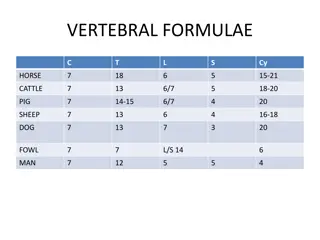
Location common uncommon Bone Femur Percentage 30 Bone Rib Tibia 15 20 Clavicle Feet & Pelvis 8 Scapula Humorous 10-20 Hands 5
Symptoms Due to mechanical effects of the lesion Nerve compression Vascular compression Reactive myositis Fracture Malignant transformation
Types Type Sessile Pedunculated Incidence Uncommon Common Location Proximal humerus and scapula Knee, hip and ankle Appearance Flat plateau like stalk producing a broad based protuberance Elongated bony stalk merging with the host bone. The hyaline cap is lobulated giving its appearance
CT Scan Allows optimal demonstration of the pathognomonic cortical and medullary continuity of the lesion and parent bone Mineralization in the cartilage cap allows a correct CT measurement Cartilage cap thickness greater than 2 cm in adults and 3 cm in growing children suggests malignant transformation
Radiological Findings - MRI Well corticated altered signal intensity lesion arising from the head of the right 3rd rib Lesion seen displacing and compressing the spinal cord towards left side
MRI scan MRI provides information about inflammation in reactive bursa formation, impingement syndrome and arterial and venous compromise This study is the method of choice for evaluating compression of the spinal cord, nerve roots and peripheral nerves
MRI Findings Lesion measures ~ 2 cm (cranio- caudal) X 1.4 cm (antero-posterior) X 1.6 cm (medio-lateral) Pedicle of the vertebral body is not imaged at this level
MRI Findings spinal cord shows intra-medullary right signal at this level and no visualization of arachnoid space compressive mylopathy changes Lesion shows bright signal on T1 and T2 sequences and low on STIR sequence
CT findings Well corticated bony lesion seen arising from medial aspect of the head of the right 3rd rib Cortex and medullary cavity of the lesion seen continuous with the cortex and medulla of the rib
CT findings The bony lesion is protruding into the adjacent neural foramina and extending into the spinal canal indentation with cortical thickening and remodelling of posterior aspect of D3 vertebral body, pedicle, lamina noted on the right side
CT Findings Rest of the visualized right 3rd rib shows mild cortical thickening Cortex and medullary cavity of the lesion seen continuous with the cortex and medulla of the rib
X-RAY Findings Lateral scalloping seen at the level of 3rd rib in the AP view
Discussion These are benign bone tumours characterised by cartilage capped bony outgrowths that project from the surface of affected bone They are mostly seen in metaphyseal portions of long bone In solitary form however the tumour tends to be less extensive and in long bones it is limited to one portion of circumference of the shaft The cartilagenous cap often has relatively acellular appearance. However, especially in those tumour of children focal areas of increased cellularity may be present
Discussion Role of radiologist is to define the lesion in the diagnosis and the effect of the lesion on the adjacent neurovascular bundle Irregularity of cartilage in a cap, a soft tissue mass projecting beyond expected confines of the lesion, flex of calcification in the soft tissue beyond the lesion and rapid growth of the lesion suggests malignant transformation Osteochondroma in areas other than knee are more likely to undergo malignant transformation Chondrosarcoma is more common in hereditary form
Discussion Dynamic gadolinium enhanced MR can be useful to differentiate benign from low grade malignant cartilaginous tumour Rib exostosis that project externally are palpable on the chest wall and internal exostosis causes pressure effect on the adjacent structure Staging for benign bone lesion given by muscular-skeletal tumour society are as follows Stage 1 inactive or static lesions Stage 2 actively growing lesions Stage 3 actively growing lesions that are locally destructive/aggressive
Discussion Exostosis of a membranous bone is extremely rare They are seen mostly in the metaphyseal portion of long bones Osteochondromas are benign developmental abnormalities in which a portion of epiphyseal growth plate cartilage becomes separated from the main epiphysis No rib swelling should be ignored since benign rib tumour is a very rare condition and can be treated conservatively unless associated with any complications or cosmetic problems
Conclusion CT is the best imaging modality for diagnosing bone lesions. Though benign rib tumour is very rare it should be evaluated cautiously when presented to OPD