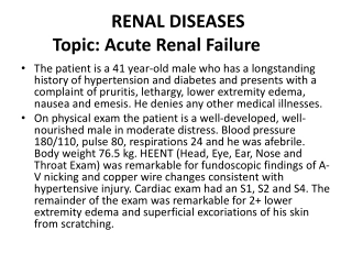
Acute renal injury in pregnancy refers to a condition where urine volume decreases significantly. Causes, pathology, and clinical features are discussed to understand this serious condition in expectant mothers.
Please find below an Image/Link to download the presentation.
The content on the website is provided AS IS for your information and personal use only. It may not be sold, licensed, or shared on other websites without obtaining consent from the author. If you encounter any issues during the download, it is possible that the publisher has removed the file from their server.
You are allowed to download the files provided on this website for personal or commercial use, subject to the condition that they are used lawfully. All files are the property of their respective owners.
The content on the website is provided AS IS for your information and personal use only. It may not be sold, licensed, or shared on other websites without obtaining consent from the author.
DEFINITION Acute renal injury (failure) is defined as a condition in which urine volume falls below 400 ml , in 24 hours or 20 ml /h Anuria :.is the absence of excretion of urine in 12 hours. Incidenc:. of acute renal failure is 1 -15,000 t0 in 2 0,000 pregnancy in industrialized countries, but in developing countries the incidence varies from 1 out of every 2000 to 1 out of every 5000 pregnancies.
Causes of Acute Renal Injury (Failure) 1-(Prerenal) in Pregnancy Early Pregnancy Late Pregnancy and Labor (1) Acute and massive hemorrhage: (1) Acute and massive hemorrhage (2) Severe dehydration (2) Abruptio placentae (3) Severe preeclampsia, eclampsia, (3) Septic abortion (4) Severe infection Other Causes in Pregnancy;. (1) Mismatched blood transfusion
(:2) Renal Renal disease, DIC, hypoperfusion, ischemia, toxins, obstetric pathology superimposed on pre-existing renal disease (interstitial nephritis (3) Postrenal (Obstructive) : Accidental ligature of the ureters during cesarean section, hysterectomy for rupture of uterus (rare)
PATHOLOGY OF ARF: Prerenal is the most common form of AKI (ARF). It is due to mild to moderate degree of renal hypoperfusion. Mild and even moderate ischemia with acute tubular necrosis are reversible. In severe ischemia, renal cortical tissue is damaged and this pathology is irreversible. ACUTE TUBULAR NECROSIS:. is the most common pathology in obstetrics. The lesion begins in loop of henle,s the renal tubules undergo ischemic degeneration and necrosis.
the Interstitial tissues become edematous. Provided the tubules have adequate blood supply, the epithelium will slowly regenerate and the renal function will usually return to normal in 1-2 weeks if it is taken care of in time. ACUTE CORTICAL NECROSIS: It is relatively uncommon and seen in abruption placenta and endotoxic shock following gram negative septicemia Usually diffuse ischemic necrosis occurs . all over the cortex.
CLINICAL FEATURES: When anuria is reversible, the clinical condition can be divided into four phases ;. 1- Incipient phase t 2-Phase of anuria 3-Phase of diuresis 4-Phase of recovery INCIPIENT PHASE: The phase is short lasting There is marked diminution in urinary output. phase of anuria is This phase lasts from a few hours to as long as 3 weeks. An urinary output is less than 400 mL in 24 hours . Initially, the patient remains alert and looks well. Gradually anorexia, vomiting and diarrhea may occur. Then the patient looks toxic; blood pressure is raised and abdomen becomes distended. Still untreated the patient becomes drowsy with dry, tongue, twitching of muscles and mental confusion .delirium followed by coma is the end result .
PHASE OF EARLY DIURESIS: In this phase, tubular reabsorptoin is delayed up to a period usually proportionate to the period of anuria. The only favorable feature is the increased excretion of urine. But the rise of potassium, sodium, creatinine, (BUN) and chloride continues and the specific gravity of the urine is still low. THE PHASE OF LATE DIURESIS: The phase is as hazardous as the previous one. The causes of diuresis are: (1) Osmotic diuresis due to high blood urea (2) Functional inadequacy of tubular reabsorption (3) Release of surplus fluid and electrolytes, particularly sodium and potassium.
PHASE OF RECOVERY: Tubular epithelium regenerates and tubular function is reestablished along with the is establishment of glomerular activity. The concentration of the electrolytes either in the plasma or in the urine gradually returns to normal values an So also the specific gravity of the urine. It may take about 1 year for restoration of full function . Differential diagnosis of acute renal failure urinalysis; perennial renal urinary Na <20meq/l >40meq/l urine osmolality FE Na <1% >2% >500 <350
MANAGEMENT OF AKI IN OBSTETRICS (Prerenal) in abruption placenta it is necessary to expand volume and improve oxygen carrying capacity and the agent of choice is packed red cells cryoprecipitate and fresh frozen plasma are used if there is a deficit in coagulation factor. in patients with sever preeclampsia this may result in pulmonary edema. These patients have increased capillary permeability and low rapidly from the intravascular in to the interstitial space. when the difference between the plasma colloid osmotic pressure (usually 21-25 mmhg )and the (usually 6-10 mmhg )is reduced to less than 3mmhg, pulmonary edema may occur.
Forced diuresis Mannitol: 100 mL of 20% mannitol is administered intravenously slowly taking at least 10 minutes. The diuresis is expected to occur within 1 hour If it fails, the infusion may be repeated after 2 hours. Frusemide Use of frusemide (Lasix) 80 120 mg intravenously, two doses at intervals of 2 hours is advocated
PHASE OF ANURIA : The principles in the management are: (1) To control the fluid balance (2) To maintain the caloric requiremen (3) To regulate the electrolyte imbalance (4) To give adequate supportive therapy (5) To prevent complications (hyperkalemia, hyperphosphatemia, hypocalcemia, metabolic acidosis).
(1) Fluid balance: In the anuric phase, water is eliminated through the extrarenal routes, i.e. lungs (approx 400 ml),skin (approx 600ml) and stool Thus, the fluid loss is approximately 1000 mL per day. . Nutrition: To meet the metabolic needs, the patient requires about 1500- 2000 Kcal/day. Protein and salts are restricted . The energy is provided mainly by carbohydrate diet . , Each 100 g of glucose gives 400 Kcal.
Complications are treated depending upon the blood values and biochemical abnormalities 1-Hyperkalemia 2- Hyperphosphatemia: 3--Metabolic acidosis Hemodialysis (Artificial kidney): Hemodialysis in pregnancy often causes wide fluctuation of blood pressure. Continuous EFM should be continued during dialysis . Patient must have at least 70 g of protein and 1.5 g of calcium daily. Hematocrit should be above 25%. Packed red cell transfusion may be given. Risk of preterm labor is high as progesterone is removed . during dialysis. Parenteral progesterone therapy is advocated in patients with dialysis.
PHASE OF DIURESIS: Fluid balance initially the fluid intake is calculated on the basis of the total amount of urinary output in previous 24 hours plus 500 mL. When the urinary output exceeds 1000 mL, oral feeding is started with high calorie low protein constituent along with fruit juice. Electrolyte balance: As there is loss of potassium the accumulated potassium isLikely to be excreted by this time potassium supplementation is require at this stage. PHASE OF RECOVERY This is recognized by general features of clinical improvement and increased amount of urine.
PROGNOSIS: Most ARF is reversible ,-in obstetrics overall mortality due to ARF is about 15% and slightly high in sepsis related ARF Renal parenchymal injury is assoc with high mortality. Prognosis of the fetus is unfavorable and there is about 50% mortality. . POSTPARTUM RENAL FAILURE (postpartum hemolytic uremic syndrome). It is a clinical condition of acute irreversible renal failure occurring within the first 6 weeks postpartum. The exact cause is still obscure. It may be due to (a) drug sensitivity (like ergot) (b) consumptive coagulopathy or (c) result of peripheral immunological mechanism. Therapy consists of hemodialysis with or without heparin OBSTRUCTIVE RENAL FAILURE;. Obstructive anuria due to ureteric ligation during c.s or hysterectomy.


")
 Renal")


")