Overview of Adrenal Imaging and Pathology
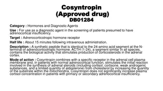
Near the top of the kidneys lie the adrenal glands secreting cortisone and adrenaline, controlling stress. They are 3 inches long, half an inch high, and have a Y-shaped endocrine structure. Imaging helps detect various adrenal pathologies like hyperplasia, infections, masses, cysts, and hemorrhages. Learn about different adrenal masses like adenomas, pheochromocytomas, carcinomas, and more through detailed imaging insights.
Download Presentation

Please find below an Image/Link to download the presentation.
The content on the website is provided AS IS for your information and personal use only. It may not be sold, licensed, or shared on other websites without obtaining consent from the author.If you encounter any issues during the download, it is possible that the publisher has removed the file from their server.
You are allowed to download the files provided on this website for personal or commercial use, subject to the condition that they are used lawfully. All files are the property of their respective owners.
The content on the website is provided AS IS for your information and personal use only. It may not be sold, licensed, or shared on other websites without obtaining consent from the author.
E N D
Presentation Transcript
SUMMARY OF ADRENALIMAGING SUMMARY OF ADRENALIMAGING ANATOMY Ad = Near /At Secrete Cortisone & Adrenaline Controlstress 1/2 inch height 3 inch length Triangle "Y" shape endocrine gland on top of kidneys. Renes = Kidney Best seen by CT & MRI Normal : Seen @ Peri-renal space Y shape By A.M.Abodahab Ass.Lecturerof radiology Sohag University
SUMMARY OF ADRENALIMAGING ADRENAL PATHOLOGY HYPERPLASIA INFECTION MASS CYST HAEMORRHAGE ADRENALMASSES Adenoma Pheochromocytoma Carcinoma Deposites Lymphoma MyeloLipoma 1- Adenoma CommonestAdrenal gland Mass Lesion < 10 Hu NECT = 90 % Adenoma Lipid Rich or Lipid poorAdenoma For confident diagnosis of LIPID RICH ADENOMA : Diagnostic = < 0 Hu in NECT * CT NECT < 10 Hu @ 60 Sec normal enhance @ 10 min significant washout MRI Out of Phase T1 SignalDrop By A.M.Abodahab Ass.Lecturerof radiology Sohag University
SUMMARY OF ADRENALIMAGING By A.M.Abodahab Ass.Lecturerof radiology Sohag University
SUMMARY OF ADRENALIMAGING Lipid Poor Adenoma : o 30 % of adenomas o @ 60 sec it enhance as other lesions o But @ 15 min Wash out > 60 % o NB. Hu assessed at center of lesion avoiding necrosis or calcification. o > 10 Hu in NECT D.D. of Adenoma: o METS o Lipid PoorAdenoma o AdrenocorticalCarcinoma o Pheochromocytoma o Adrenal Graneulomatus disease Conn's Disease: -Excess mineralocorticoid production - 1 M : 2 F - 30 : 50 Y o Causes : Adenoma 70 % Hyperplasia or Carcinoma o C.P.: Hypertension / HypoKalimeia Cushing Syndrome: o Excess Gluco-corticoidproduction o o Causes: Pituitary adenoma Adrenal Adenoma 20% Adrenocortical Carcinoma 10% Adrenal Hyperplasia MainC.P .: - D.M. II -Hypertension -Hypercholest. Abd pain By A.M.Abodahab Ass.Lecturerof radiology Sohag University
SUMMARY OF ADRENALIMAGING 2-AdrenalHyperplasia Causes : - Pituitary Adenoma over ACTH - S t r e s s - AgingProcess Bilateral AdrenalHy perplasia Adrenal Hyperplasia Active in PETCT D.D. Adrenal Hyperplasia : o Adenoma o Haemorrhage o Aging 3- PHEOCHROMOCYTOMA IV CONTRAST IS CONTRAINDICATED HYPERTENSIVE CRISIS mayOCCUR Age: 4th : 6th d e c a d e From Adrenal M e d u l l a NON SOECIFIC IMAGING FINDING Roleof 10 Lab Diagnosis : o Elevated Catecholamines : In Urine 97 % sensitivity In Plasma 99% sensitivity Adrenal Mass + VM Vanillyl Mandelic Acid in urine + Hypertension = Mostly PHEOCHROMOCYTOMA By A.M.Abodahab Ass.Lecturerof radiology Sohag University
SUMMARY OF ADRENALIMAGING 4-Adeno-corticalCarcinoma Arise from Adrenal Cortex Small amount of hormones "No symptoms" Large "> 6 cm" up to 20 cm Heterogenous enhancement Calcification 30 % of cases Can invade adjacent structures 5-AdrenalDeposits Source : Lungs, Breast & Skin .& may O t h e r s NON SOECIFIC IMAGING FINDING +/- Signs of malignancy : Known primary + Metabolic active lesion 6-AdrenalLymphoma Unilateral or Bilateral + Lymphadenopathy 7-AdrenalMyelolipoma Rare (FAT CONTAINING) Benign , No malignant transformation Adults usually Incidentally Discovered Non Functioning usually Complication : Hemorrhage By A.M.Abodahab Ass.Lecturerof radiology Sohag University
SUMMARY OF ADRENALIMAGING SmallAdrenal Myelolipoma ADRENALCYST Rare F 2 : 1 M 30 : 50 y C.P .: o Usually asymptomatic Complecations: -Rupture -Haemorrhage -Infection CYST = NO CONTRAST Fluidsignal CECT Cyst is not enhancing ADRENAL HYDATID CYST By A.M.Abodahab Ass.Lecturerof radiology Sohag University
SUMMARY OF ADRENALIMAGING ADRENAL HAEMORRHAGE Blood Density oval Lesion 90% Rt Between live & spine If + Primary Mets to be considered ADRENAL INFECTION Bilateral enlarged + Ca +ve skin Tubreculin test Highly suggestive of T B Diagnostic BILATERAL ADRENAL CALCIFICATION Previous Haemorhage Familial Medtrainian Fever Incidental Finding By A.M.Abodahab Ass.Lecturerof radiology Sohag University SOURCES : Lecture of Prof. MamdouhMahfouz JULY2018 http://www.radiologyassistant.nl/en/p421aee


