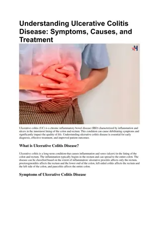
Overview of Inflammatory Bowel Disease (IBD) and Ulcerative Colitis
Inflammatory bowel disease (IBD) encompasses ulcerative colitis (UC) and Crohn's disease (CD), with varying epidemiology, clinical features, and pathogenesis. Understand the manifestations, factors affecting susceptibility, and distribution of these conditions, particularly UC which involves distinct patterns like ulcerative proctitis, proctosigmoiditis, left-sided colitis, and pancolitis. Genetic and environmental elements play crucial roles in IBD development. Explore the complexities and nuances of IBD for comprehensive insights.

Download Presentation

Please find below an Image/Link to download the presentation.
The content on the website is provided AS IS for your information and personal use only. It may not be sold, licensed, or shared on other websites without obtaining consent from the author.If you encounter any issues during the download, it is possible that the publisher has removed the file from their server.
You are allowed to download the files provided on this website for personal or commercial use, subject to the condition that they are used lawfully. All files are the property of their respective owners.
The content on the website is provided AS IS for your information and personal use only. It may not be sold, licensed, or shared on other websites without obtaining consent from the author.
E N D
Presentation Transcript
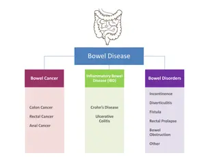
Inflammatory bowel disease (IBD) is comprised of two major disorders: Ulcerative colitis (UC). Crohn's disease (CD). These disorders have both distinct and overlapping pathologic and clinical characteristics.
Epidemiology IBD is more common in the West, but the incidence is increasing in the developing countries including Saudi Arabia. IBD can present at any age: The peak :15 - 30 years. A second peak 50
Inflammatory bowel disease (IBD) is comprised of two major disorders: Ulcerative colitis (UC) Crohn's disease (CD). These disorders have distinct pathologic and clinical characteristics, but their pathogenesis remains poorly understood Medium High Low
HOST FACTORS Genetic factors: NOD2/CARD15 ENVIRONMENTAL FACTORS Smoking: Appendectomy: protect UC Diet
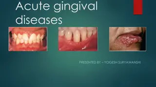
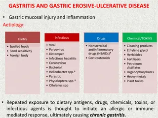
Ulcerative colitis Ulcerative colitis is characterized by recurring episodes of inflammation limited to the mucosal layer of the colon. Start rectum then extend proximally.
Ulcerative proctitis :rectum. Ulcerative proctosigmoiditis :rectum and sigmoid colon. Left-sided colitis: disease that extends beyond the rectum and as far proximally as the splenic flexure. Extensive colitis :beyond the splenic flexure. Pancolitis: whole colon
40-50% of patients have disease limited to the rectum and rectosigmoid 30-40% of patients have disease extending beyond the sigmoid 20% of patients have pancolitis
The major symptoms of UC are: - diarrhea - rectal bleeding - tenesmus - passage of mucus - crampy abdominal pain
Patients with proctitis usually pass fresh blood or blood- stained mucus either mixed with stool or streaked onto the surface of normal or hard stool When the disease extends beyond the rectum, blood is usually mixed with stool or grossly bloody diarrhea may be noted When the disease is severe, patients pass a liquid stool containing blood, pus, fecal matter Other symptoms in moderate to severe disease include: anorexia, nausea, vomitting, fever, weight loss
DIAGNOSIS No single modalities is enough for Diagnosis. Combination of Clinical picture, laboratory, Endoscopy, pathology.
Colonoscopy The vascular markings are lost, petechiae, exudates, touch friability, and frank hemorrhage may be present. Colonic involvement is continuous in ulcerative colitis, in contrast to the patchy nature of Crohn's disease.
Pathology: crypt abscesses. chronic changes including branching of crypts, atrophy of glands, and loss of mucin in goblet cells
Normal Video Colonoscopy. Showing characteristic triangular folds of the transverse colon. With the patient in left lateral decubitus position, a long, flexible, lighted viewing tube (colonoscope) is inserted through the rectum into the colon. The scope is advanced and maneuvered while the lumen and walls of the colon are visualized by projections onto a monitor screen. The colonoscope has channels through which instruments can be passed in order to perform biopsies, remove polyps, or cauterize bleeding. Air, water, and suction can be applied to help provide a clearer visual field for inspection.
Ulcerative colitis - complication Hemorrhage Perforation Toxic megacolon (transverse colon with a diameter of more than 5,0 cm to 6,0 cm with loss of haustration) Colon cancer
managements Goals of therapy Induce and maintain remission. Ameliorate symptoms Improve pts quality of life Adequate nutrition Prevent complication of both the disease and medications
managements Role out infection 5 ASA therapy: Oral Rectal Corticosteroids: Systemic: Prednisolone Local acting: enema. Immunomodulators : Azithyoprine Methotrexate Anti TNF therapy
Surgery: Severe attacks that fail to respond to medical therapy. Complications of a severe attack (e.g., perforation, acute dilatation). Chronic continuous disease with an impaired quality of life. Dysplasia or carcinoma.
CD Crohn's disease (CD) is a disorder of uncertain etiology that is characterized by transmural inflammation of the gastrointestinal tract. CD may involve the entire gastrointestinal tract from mouth to the perianal area. 80% Small bowel. 50 % ileocolitis. 20 % colon. 30% perianal disease. UGI < 5 %
CLINICAL MANIFESTATIONS Fatigue. Diarrhea. Abdominal pain. Weight loss. Fever.
Phlegmon/abscess : walled off inflammatory mass without bacterial infection Fistulas : Fistulas are tracts or communications that connect two epithelial-lined organs. Enterovesical Enterocutaneous Enteroenteric enterovaginal Perianal disease
Severe oral involvement aphthous ulcers. Esophageal involvement odynophagia and dysphagia. Gastroduodenal CD upper abdominal pain and symptoms of gastric outlet obstruction. Gallstones
Extraintestinal manifestations CD and ulcerative colitis share a number of extraintestinal manifestations Arthritis Eye involvement uveitis, iritis, and episcleritis Skin disorders. Others: Primary sclerosing cholangitis, Venous and arterial thromboembolism ,Renal stones ,Bone loss and osteoporosis ,Vitamin B12 deficiency
DIAGNOSIS The diagnosis of CD is usually established with endoscopic findings or imaging studies in a patient with a compatible clinical history.
Colonoscopy: Endoscopic features include focal ulcerations adjacent to areas of normal appearing mucosa along with polypoid mucosal changes that give a cobblestone Wireless capsule endoscopy
Normal Video Colonoscopy. Showing characteristic triangular folds of the transverse colon. With the patient in left lateral decubitus position, a long, flexible, lighted viewing tube (colonoscope) is inserted through the rectum into the colon. The scope is advanced and maneuvered while the lumen and walls of the colon are visualized by projections onto a monitor screen. The colonoscope has channels through which instruments can be passed in order to perform biopsies, remove polyps, or cauterize bleeding. Air, water, and suction can be applied to help provide a clearer visual field for inspection.
Imaging studies small bowel follow through (SBFT) computed tomography: CTS or CT enterography Magnetic resonance imaging (MRI) or MR enterography
Serologic markers Inflamatory marker : ERS, CRP Antibody tests : Antineutrophil cytoplasmic antibodies (pANCA) Anti-Saccharomyces cerevisiae antibodies (ASCA) Stool markers fecal calprotectin
Goals of therapy Induce and maintain remission. Ameliorate symptoms Improve pts quality of life Adequate nutrition Prevent complication of both the disease and medications
Management Role out infection Corticosteroids: Systemic: Prednisolone Local acting: Budosonide. Immunomodulators : azithyoprine Methotrexate
Anti TNF therapy Surgery Obstruction, severe perianal disease unresponsive to medical therapy, difficult fistulas, major bleeding, severe disability
Distinguishing characteristics of CD and UC Feature Location CD UC colon SB or colon Anatomic distribution Rectal involvement Gross bleeding Peri-anal disease Fistulization Granulomas Skip lesions Continuous Rectal spare Involved in >90% Only 25% 1/3 Yes 30% Universal Rare No No
Endoscopic features of CD and UC Feature Discontinuous Mucosal involvement Aphthous ulcers CD UC Continuous Common Rare Surrounding mucosa Longitudinal ulcer Cobble stoning Mucosal friability Vascular pattern Relatively normal Common In severe cases Uncommon Normal Abnormal Rare No Common distorted
Pathologic features of CD and UC Feature CD Yes UC Transmural inflammation Uncommon Granulomas 30% No Fissures Fibrosis Common Common Common Rare No Submucosal inflammation Uncommon

 is comprised of")

 is comprised of two")
 is comprised of two")