Periodontal Diseases and Radiographic Assessment
Learn about periodontal diseases such as gingivitis and periodontitis, their etiology, clinical features, role of radiographs in assessment, and anatomical considerations. Explore how radiographs aid in evaluating bone status, identifying contributing factors, and understanding periodontal conditions while acknowledging their limitations.

Download Presentation

Please find below an Image/Link to download the presentation.
The content on the website is provided AS IS for your information and personal use only. It may not be sold, licensed, or shared on other websites without obtaining consent from the author. If you encounter any issues during the download, it is possible that the publisher has removed the file from their server.
You are allowed to download the files provided on this website for personal or commercial use, subject to the condition that they are used lawfully. All files are the property of their respective owners.
The content on the website is provided AS IS for your information and personal use only. It may not be sold, licensed, or shared on other websites without obtaining consent from the author.
E N D
Presentation Transcript
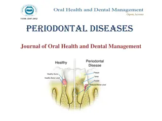
Most common is gingivitis and periodontitis Gingivitis is a soft tissue inflammation involving the gingiva surrounding teeth Periodontitis entails loss of soft tissue attachment and supporting bone of involved teeth
Factors affecting and etiology: Diabetes Smoking Genetic predispostion Subclassified as: chronic, aggressive, necrotizing, those related to abscess formation and those in conjunction with endodontic lesions
Clinical features Gingivitis: gingival swelling, edema and erythema Progression of the disease leads to pocket formation Other signs include bleeding, purulent exudate, edema, resorption of alveolar crest and tooth mobility
Role of radiographs r/g play an integral role in assessment of periodontal disease They provide unique information about the status of periodontium and a permanent record of condition of bone throughout the course of disease They aid in identifying the extent of destruction of alveolar bone, local contributing factors and features of periodontium that influence the prognosis
R/G assessment of periodontal conditions Help in evaluation of amount of bone present Condition of alveolar crests Bone loss in furcation areas Width of PDL space Local initiating factors that cause or intensify periodontal disease 1. Calculus 2. Poorly contoured or overextended restorations Root length and morphology and crown to root ratio
Anatomic considerations 1. Position of maxillary sinus in relation to periodontal deformity 2. Missing, supernumerary, or impacted teeth Pathologic considerations 1. Caries 2. Periapical lesions 3. Root resorption
Limitations of radiographs R/G provide a 2 dimensional view of a 3-dimensional situation R/G typically show less severe bone destruction than is actually present They do not demonstrate the soft tissue to hard tissue relationships and thus provide no information about depth of soft tissue pockets Bone level is often measured from CE junction. This reference point is not valid in situations in which either overeruption or severe attrition with passive eruption exists
Bitewing images more accurately record the distance b/w the CEJ and the crest of interradicular alveolar bone IOPA view, splly the posterior maxilla may present a distorted view of relationship b/w teeth and height of alveolar bone Teeth will be dipicted in their correct positions relative to alveolar process if No overlapping of proximal contacts b/w crowns No overlapping of roots of adjacent teeth Overlapping of buccal and lingual cusps of molars
Special considerations and techniques It is useful to superimpose fine wire grids when exposing R/g to aid the measurement of relative bone height These grids form 1mm squares that allow quantitative measurement of position of alveolar bone with respect to dentition Subtraction radiography: it allows better detection of small amounts of bone loss b/w radiographs made at different times
Normal anatomy A thin layer of opaque cortical bone often covers the alveoalr crest Height of the crest lies at a level approx 1 to 1.5mm below the level of CEJ of adjacent teeth b/w anterior teeth, crest is usually pointed and has a dense cortex In anteriors, crest lies within 1 to 1.5mm of a line connecting the adjacent CEJs Alveolar crest is in line with lamina dura of adjacent teeth In posteriors, junction b/w crest and lamina dura forms a sharp line next to tooth root
General R/G features of periodontal disease Changes can be divided into: Changes in morphology of supporting alveolar bone Changes to internal density and trabecular pattern
Mild periodontitis Early lesions appear as areas of localized erosions of interproximal alveolar bone crest Anterior regions show blunting of crests and slight loss of alveolar bone height Posterior regions may also show a loss of sharp angle b/w lamina dura and crest In early periodontal disease, angle may lose its normal cortical surface and appear rounded off, having an irregular and diffuse border
Moderate periodontitis the destruction of alveolar bone extends beyond early changes in the alveolar crest and may induce a variety of defects in the morphology of alveolar crest The patterns of bone loss are divided as: 1. Localized buccal or lingual cortical plate loss 2. Gen horizontal bone loss in a region 3. Localized vertical defects involving just one or two teeth
Buccal or lingual cortical plate loss Buccal or lingual cortical plate adjacent to the teeth may resorb This is indicated by an increase in r lucency of root of teeth near alveolar crest It is semicircular shadow with apex of r lucency directed apically in relation to tooth
Horizontal bone loss Loss in height of alveolar bone around multiple teeth Crest is still horizontal but positioned apically more than a few mm from the line of CEJ It may be mild, moderate or severe, depending on extent Mild bone loss is 1mm of attachment loss Moderate is greater than 1mm upto the midpoint of length of roots or to the furcation level of molars Severe loss is anything beyond that point and often has evidence of furcation involvement of multirooted tooth
Vertical osseous defects Are localized to one or two teeth Crest displays an oblique angulation to the line of CEJ in the area of involved teeth Are divided into 2 primary types: Interproximal crater is a 2 walled, trough like depression that forms in the crest of interdental bone b/w adjacent teeth Infrabony defect is a vertical deformity within bone that extends apically along the root from the alveolar crest
Infra bony defect is described as 3 walled (surrounded by 3 bony walls) when both cortical plates remain It is 2 walled when one of these plates has been resorbed One walled when both plates have been lost
Surrounding internal bone changes R lucent change reflects loss of density and no of trabeculae, and trabeculae appear faint, commonly seen in early or acute lesions The apparent reformation of bone when acute inflammation resolves, trabeculae remineralize Sclerotic bone reaction appears r opaque as a result of deposition of bone on existing trabeculae at the expense of the marrow, resulting in thicker trabeculae that may eventually be so dense as to appear as an amorphous r opaque mass
Severe periodontitis The bone loss is so extensive that the remaining teeth show excessive mobility and drifting coz of inadequate support Extensive horizontal bone loss or extensive vertical osseous defects may be present
Osseous deformities in the furcations Progressive periodontal disease and its associated bone loss may extend into the furcations As bone resorption extends down the side of multirooted teeth, it can reach the level of furcation and beyond Widening of PDL space at apex of interradicular bony crest of furcation is strong evidence that periodontal disease process involves furcation Bony defect may involve either buccal or lingual cortical plate and extend under roof of furcation. Here the defect does not involve other cortical plate and appear more irregular and r lucent than adjacent normal bone
If crestal bone is below furcation but disease process has not extended into interradicular bone, the width of PDL space appears normal Furcation defects involve maxillary molars about 3 times as often as mandibular molars Loss of interradicular bone in furcation of upper molar may originate from buccal, mesial or distal sueface of teeth Occassionally Bone loss appears as inverted J shadow with the hook of J extending into trifurcation
Periodontal abscess Is a rapidly progressing, destructive lesion that usually originates in a deep soft tissue pocket Occurs when coronal portion of the pocket becomes occluded or when foreign material becomes lodged b/w a tooth and gingiva Pain and swelling are present in the region If the lesion persists, a r lucent region appears, often superimposed over the root of a tooth A bridge of bone may be present over the coronal aspect of the lesion, separting it from the crest of the alveolar ridge
Aggressive periodontitis Refers to periodontal disease with an aggressive and rapid nature that occurs in pts under 30 yrs of age Severity of disease appears to be an exuberant reaction to a minimum amount of plaque accumulation and may result in early tooth loss Classified as: 1. localized aggressive periodontitis 2. Generalized aggressive periodontitis
Clinical presentation Localized aggressive periodontitisis associated with attachment loss involving the incisors and first molars Commences around puberty and bone loss is rapid, upto 3-4 times that seen in chronic periodontitis Few signs of soft tissue inflammation or plaque accumulation present despite presence of deep bony pockets Pt will present with drifting and mobile incisors and early loss of 1stmolars
Generalized aggressive periodontitis: involves a variable no of teeth from a least 3 to all of the dentition Usually affects under 30 yrs of age Gingiva appear normal as in localized form or may present with exuberant inflammatory response
Radiographic presentation In localized form is typically vertical Maxillary teeth are involved more than mandibular teeth and a strong left-right symmetry is common Gen form can involve several teeth or all dentition and the rapid bone loss may be of vertical or angular or horizontal pattern