Pulmonary Emphysema: Pathophysiological Changes & Consequences
Pulmonary emphysema, a result of smoking, leads to chronic lung infection, obstruction of airways, alveolar destruction, and a barrel chest. The condition progresses slowly, causing severe air hunger and ultimately death. Pneumonia, on the other hand, involves lung inflammation and alveolar fluid build-up, compromising gas exchange and leading to severe consequences if not treated promptly.
Download Presentation

Please find below an Image/Link to download the presentation.
The content on the website is provided AS IS for your information and personal use only. It may not be sold, licensed, or shared on other websites without obtaining consent from the author.If you encounter any issues during the download, it is possible that the publisher has removed the file from their server.
You are allowed to download the files provided on this website for personal or commercial use, subject to the condition that they are used lawfully. All files are the property of their respective owners.
The content on the website is provided AS IS for your information and personal use only. It may not be sold, licensed, or shared on other websites without obtaining consent from the author.
E N D
Presentation Transcript
APPLIED ASPECTS OF RESPIRATION APPLIED ASPECTS OF RESPIRATION II. II. DR. M. M. KHAN PROF. & HEAD DEPTT. OF PHYSIOLOGY
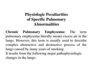
PATHOPHYSIOLOGY OF SPECIFIC PULMONARY ABNORMALITIES (1). CHRONIC PULMONARY EMPHYSEMA The term pulmonary emphysema literally means excess air in the lungs. However, this term is usually used to describe a complex obstructive and destructive process of the lungs caused by many years of smoking. It results into following major pathophysiological changes in the lungs : 1. Chronic infection, caused by inhaling smoke or other substances that irritate the bronchi and bronchioles. The chronic infection seriously deranges the normal protective mechanisms of the airways, including partial paralysis of the cilia of the respiratory epithelium, an effect caused by nicotine. As a result, mucus cannot be moved easily out of the passageways.
2. The infection, excess mucus, and inflammatory edema of the bronchiolar epithelium together cause chronic obstruction of many of the smaller airways. 3. The obstruction of the airways makes it especially difficult to expire, thus causing entrapment of air in the alveoli and overstretching them. This effect, combined with the lung infection, causes marked destruction of as much as 50 to 80 percent of the alveolar walls. 4. over a period of years, the chest cage becomes permanently enlarged, causing a barrel chest, . Thus lungs become voluminous with showing more vertical diameter of lungs in x ray.
5. Chronic emphysema usually progresses slowly over many years. Both hypoxia and hypercapnia develop because of hypoventilation of many alveoli plus loss of alveolar walls. The net result of all these effects is severe, prolonged, devastating air hunger that can last for years until the hypoxia and hypercapnia cause death a high penalty to pay for smoking.
(2). PNEUMONIA LUNG INFLAMMATION AND FLUID IN ALVEOLI The term pneumonia includes any inflammatory condition of the lung in which some or all of the alveoli are filled with fluid and blood cells. A common type of pneumonia is bacterial pneumonia, caused most frequently by pneumococci. This disease begins with infection in the alveoli; the pulmonary membrane becomes inflamed and highly porous so that fluid and even red and white blood cells leak out of the blood into the alveoli. Eventually, large areas of the lungs, sometimes whole lobes or even a whole lung, become consolidated, which means that they are filled with fluid and cellular ndebris
In persons with pneumonia, the gas exchange functions of the lungs decline in different stages of the disease. In early stages, the pneumonia process might well be localized to onlyone lung, with alveolar ventilation. This condition causes two major pulmonary abnormalities: (1) reduction in the total available surface area of the respiratory membrane. (2) a decreased ventilation-perfusion ratio. Both of these effects cause hypoxemia (low blood O2) and hypercapnia (high blood CO2).
(3.) ATELECTASIS COLLAPSE OF THE ALVEOLI : Atelectasis means collapse of the alveoli. It can occur in localized areas of a lung or in an entire lung. Common causes of atelectasis are : (1) total obstruction of the airway , or (2) lack of surfactant in the fluids lining the alveoli. 1. Airway obstruction causes lung collapse. The airway obstruction type of atelectasis usually results from : (a) blockage of many small bronchi with mucus, or (b) obstruction of a major bronchus by either a large mucus plug or some solid object such as a tumor. The air entrapped beyond the block is absorbed within minutes to hours by the blood flowing in the pulmonary capillaries.
2. Lack of Surfactants as a Cause of Lung Collapse. Surfactants are secreted by special alveolar epithelial cells type 2 , into the fluids that coat the inside surface of the alveoli. The surfactants in turn decrease the surface tension in the alveoli 2 to 10 - fold, which normally plays a major role in preventing alveolar collapse. However, in several conditions, such as in hyaline membrane disease (also called respiratory distress syndrome), which often occurs in premature newborn babies, the quantity of surfactants secreted by the alveoli is so greatly depressed that the surface tension of the alveolar fluid becomes several times of normal. This situation causes a serious tendency for the lungs of these babies to collapse or to become filled with fluid . Many of these infants die of suffocation when large portions of the lungs become atelectatic.
(4.) BRONCHIAL ASTHMA SPASMODIC CONTRACTION OF SMOOTH MUSCLES IN BRONCHIOLES : Asthma is characterized by spastic contraction of the smooth muscleS in the bronchioles, which partially obstructs the bronchioles and causes extremely difficult breathing. It occurs in 3 to 5 percent of all people at some time in life. The usual cause of asthma is contractile hypersensitivity of the bronchioles in response to foreign substances in the air. In about 70 percent of patients younger than age 30 years, the asthma is caused by allergic hypersensitivity, especially sensitivity to plant pollens. In older people, the cause is almost always hypersensitivity to non-allergenic types of irritants in the air, such as irritants in smog.
The bronchiolar diameter becomes more reduced during expiration than during inspiration in persons with asthma. As a result of bronchiolar collapse during expiratory effort that compresses the outsides of the bronchioles. Because the bronchioles of the asthmatic lungs are already partially occluded, further occlusion resulting from the external pressure creates especially severe obstruction during expiration. That is why , the asthmatic person often can inspire quite adequately but has great difficulty in expiring. Also, all of these together result in dyspnea, or, breathlessness, or , air hunger . The typical allergic person tends to form abnormally large amounts of IgE antibodies, and these antibodies cause allergic reactions.
(5). TUBERCULOSIS In tuberculosis, the tubercle bacilli cause a peculiar tissue reaction in the lungs, including : (1) invasion of the infected tissue by macrophages and (2) walling off of the lesion by fibrous tissue to form the so-called tubercle. This walling-off process helps to limit further transmission of the tubercle bacilli in the lungs and therefore is part of the protective process against extension of the infection. However, in about 3 percent of all people in whom tuberculosis develops, if the disease is not treated, the walling off process fails and tubercle bacilli spread throughout the lungs, often causing extreme destruction of lung tissue with formation of large abscess cavities.
(6). (6). DISEASES OF PLEURA DISEASES OF PLEURA 1. PLEURAL EFFUSION : Inflammatory, or , Exudative fluid in pleural cavity , e.g. in Tuberculosis, Pneumonia. 2. HYDROTHORAX : Non inflammatory, or, Transudative fluid in pleural cavity , e.g. in Heart failure , etc. 3. PNEUMOTHORAX : Air in pleural cavity. 4. HAEMOTHORAX : Blood in pleural cavity . 5. CHYLOTHRAX : Lymph in pleural cavity . 6. PLEURISY : Thickened & inflammatory pleura, e.g. Tuberculosis


