Pulmonary tuberculosis is a chronic, often deadly infectious disease caused by Mycobacterium tuberculosis. It involves a chronic inflammatory response and tissue destruction in the lungs. Learn about its transmission, pathogenesis, and cellular players.
Please find below an Image/Link to download the presentation.
The content on the website is provided AS IS for your information and personal use only. It may not be sold, licensed, or shared on other websites without obtaining consent from the author. If you encounter any issues during the download, it is possible that the publisher has removed the file from their server.
You are allowed to download the files provided on this website for personal or commercial use, subject to the condition that they are used lawfully. All files are the property of their respective owners.
The content on the website is provided AS IS for your information and personal use only. It may not be sold, licensed, or shared on other websites without obtaining consent from the author.
Pulmonary Tuberculosis Tuberculosis sa detaiverbba(TB roftubercle bacillus ro Tuberculosis) is a common and often deadly infectious disease caused by mycobacteria, mainly Mycobacterium tuberculosis . eht skcatta yllausu sisolucrebuT sa( sgnul yranomlup BT .
Definition Chronic inflammatory response of prolonged duration (weeks,months,years) provoked by the persistence of the causative organism Mycobacterium tuberculosis accompanied by tissue destruction and repair of lung tissue.
TB Transmission TB is spread through tiny drops sprayed into the air when an infected person coughs, sneezes, or speaks, or another person breathes the air into their lungs containing the TB bacteria.
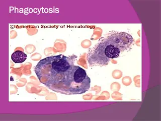
Pathogenesis :The Players (mononuclear phagocyte system) Macrophages Scattered all over (microglia, Kupffer cells, sinus histiocytes, alveolar macrophages, etc.) Circulate as monocytes and reach site of injury within 24 48 hrs and transform Become activated by T cell-derived cytokines, endotoxins, and other products of inflammation
The Players. T and B lymphocytes Antigen-activated (via macrophages and dendritic cells) Release macrophage-activating cytokines (in turn, macrophages release lymphocyte-activating cytokines until inflammatory stimulus is removed) Plasma cells Terminally differentiated B cells Produce antibodies
The dominant cellular player in pulmonary tuberculosis is the tissue macrophage Blood monocyte migrate into tissue within 48 hours after injury Tissue macrophage (RES) Kupffer cell (liver) Microglia (CNS) Histiocytes (spleen) Alveolar macs (lung) and differentiate It is joined by lymphocytes and plasma cells, however mast cells and eosinophils are as well involved in chronic allergic diseases Plasma cell Lymphocyte
Pathogenesis A-Primary Pulmonary Tuberculosis (0-3 weeks): inhalation of Mycobacteria---enter alveolar macrophages---replication in phagosome--- bacteremia---seeding of multiple sites
Primary Tuberculosis Develops in unexposed, unsensitized persons Mostly(95%) asymptomatic; Infection contained In 5 % symptomatic; Primary progressive tuberculosis Source of organism; Exogenous Difficult to diagnose
Primary Tuberculosis Presentation : Lung consolidation; Lower and middle lobes Hilar lymphadenopathy Pleural effusion In majority organism remain dormant In severe immunosupression - TB Meningitis - Miliary Tuberculosis
Secondary Tuberculosis Arises in previously sensitized host Many years after primary infection Reactivation of latent infection or overwhelming exogenous infection Involves apex of upper lobes Cavitation more prominant
Morphology A-Primary Tuberculosis : Ghon complex a bout(1 cm ) area of gray-white inflammatory consolidation develops (called Ghon focus). The center of this focus undergoes caseous necrosis. - subpleural parenchymal lesion (lower part of upper lobe) - hilar lymphadenopathy Progressive fibrosis Calcification
Histology : Epithelioid cell granulomas epithelioid cells lymphocytes plasma cells multinucleated langhan s type giant cells with or without central caseation
Granulomatous Inflammation Clusters of T cell-activated macrophages, which engulf and surround indigestible foreign bodies (mycobacteria, H. capsulatum, silica, suture material) Resemble squamous cells, therefore called epithelioid granulomas
Morphology B Secondary Tuberculosis : Focus of consolidation, < 2 cm Within 1-2 cm of apical pleura Histology : Caseating granulomas
Morphology c Miliary Pulmonary Tuberculosis : Organisms draining through lymphatics enter venous blood and circulate back into the lung and involving most of the parenchymal tissue. Microscopic or small visible yellowish white foci resembling millet seeds Coalesce to form larger cavity
Acute respiratory distress syndrome (ARDS) Previously known as respiratory distress syndrome (RDS )or adult respiratory distress syndrome:is a severe, life-threatening medical condition characterized by widespread inflammation in the lungs. ARDS is a disease of the microscopic air sacs of the lungs called (alveoli) that leads to decreased exchange of oxygen and carbon dioxide. ARDS is associated with several pathologic changes: the release of inflammatory chemicals, breakdown of the cells lining the lung's blood vessels and surfactant loss leading to increased surface tension and fluid accumulation in the lung.
Surfactant a mixture of phospholipids and lipoproteins , is secreted by lung cells. The air-fluid interface of the lining the alveoli of the lung (where the exchange of oxygen and CO2 occurs) exerts large forces that cause the alveoli to close if surfactant is deficient. Lung compliance is decreased, and the work of inflating the stiff lungs is increased.
Cystic fibrosis (CF) is a multisystem disease affecting the lungs, digestive system, sweat glands, and the reproductive tract. Patients with CF have abnormal transport of chloride and sodium across secretory epithelia, resulting in thickened, viscous secretions in the bronchi, biliary tract, pancreas, intestines, and reproductive
Although the disease is systemic, progressive lung disease continues to be the major cause of morbidity and mortality for most patients. Over a highly variable time course ranging from months to decades after birth, individuals eventually develop chronic infection of the respiratory tract with a characteristic array of bacterial flora, leading to progressive respiratory insufficiency and eventual respiratory failure
Etiology Cystic fibrosis is an autosomal recessive disease caused by defects in the CFTRgene, which encodes for a protein that functions as a chloride channel, and also regulates the flow of other ions across the apical surface of epithelial cells.The CF locus was localized through linkage analysis to the long arm of human chromosome 7, band q31
Pathophysiology Cystic fibrosis is caused by defects in the cystic fibrosis gene, which codes for a protein transmembrane conductance regulator (CFTR) that functions as a chloride channel and is regulated by cyclic adenosine monophosphate (cAMP). Mutations in the CFTR gene result in abnormalities of cAMP-regulated chloride transport across epithelial cells on mucosal surfaces
Defective CFTR results in decreased secretion of chloride and increased reabsorption of sodium and water across epithelial cells. The resultant reduced height of epithelial lining fluid and decreased hydration of mucus results in mucus that is stickier to bacteria, which promotes infection and inflammation. Secretions in the respiratory tract, pancreas, GI tract, sweat glands, and other exocrine tissues have increased viscosity, which makes them difficult to clear. Most patients with cystic fibrosis have severe chronic lung disease and exocrine pancreatic insufficiency. Additional manifestations include the following: Nasal polyposis Pansinusitis Rectal prolapse Chronic diarrhea Pancreatitis Cholelithiasis Cirrhosis or other forms of hepatic dysfunction
Cysic fibrosis Lung disease Most deaths associated with cystic fibrosis result from progressive and end-stage lung disease. In individuals with cystic fibrosis, the lungs are normal in utero, at birth, and after birth, before the onset of infection and inflammation .Shortly after birth, many persons with cystic fibrosis acquire a lung infection, which incites an inflammatory response. Infection becomes established with a distinctive bacterial flora. A repeating cycle of infection and neutrophilic inflammation develops.
Pulmonary Embolism is blockage of artery of lungs due to blood clots which might have travelled from mostly legs or other parts of body to lungs through bloodstream. As pulmonary Embolism derives blood clot in legs called deep vein thrombosis, it is referred as venous thromboembolism. Symptoms include shortness of breath, chest pain, cyanosis and rapid heart rate.


")