Renal Tubular Acidosis: Types and Causes

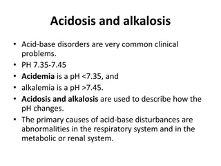
Renal tubular acidosis (RTA) is a condition where the kidneys fail to excrete enough acid, affecting pH balance. Learn about the types of RTA, renal handling of acid-base status, and alkalosis causes.

Download Presentation

Please find below an Image/Link to download the presentation.
The content on the website is provided AS IS for your information and personal use only. It may not be sold, licensed, or shared on other websites without obtaining consent from the author.If you encounter any issues during the download, it is possible that the publisher has removed the file from their server.
You are allowed to download the files provided on this website for personal or commercial use, subject to the condition that they are used lawfully. All files are the property of their respective owners.
The content on the website is provided AS IS for your information and personal use only. It may not be sold, licensed, or shared on other websites without obtaining consent from the author.
E N D
Presentation Transcript
Acid Acid- -Base Disorders & Base Disorders & Blood Gas Analysis Blood Gas Analysis APRIL 2018 CLINICAL PATHOLOGY FELLOWSHIP PROGRAM
Renal Handling of acid Renal Handling of acid- -base status: to maintain a normal amount of both H to maintain a normal amount of both H+ +and HCO base status: How does the kidney help How does the kidney help and HCO3 3- -? ? The renal glomeruli filter H+ and any acid anions present The Proximal tubules reclaim bicarb with high effectiveness (70-80%) Filtration of H+ and A- Aldosterone: exchange Na+ for H+ &K+ Reabsorption of HCO3- H+ secretion The distal tubules and collecting ducts secretes H+ as NH4+ Aldosterone acts on the cortical collecting duct to reabsorb Na+ in exchange for K+ and/or H+
Renal Tubular Acidosis (RTA) Renal Tubular Acidosis (RTA) In RTA, the kidneys inappropriately fail to excrete the amount of acid needed to maintain a normal pH. The renal defect could occur at different points of the nephron. Glomerular damage loss of entire nephrons and reduction of the number of tubules available for excretion of H+ and reabsorption of bicarb.
Types of Renal tubular acidosis Type Site of defect I Distal tubule II Proximal tubule Defective reabsorption of many other substances IV Distal tubule Aldosterone deficiency or resistance, high K+ Associated features Kidney stones, low K+ Hyperchloremic acidosis with hyperkalemia, similar to type IV RTA is seen in the majority of patients with mild to moderate chronic renal failure, even if due to glomerular disease.
ALKALOSIS ALKALOSIS Causes: 1. increased excretion of acid: the most common mechanism 2. decreased excretion of base: a common contributor to alkalosis, but rarely the only cause. Also called: Contraction alkalosis or Chloride-responsive alkalosis 3. increase addition of alkali: the body does not naturally manufacture base as a metabolic end product, all cases are due to alkali administration i.e. exogenous alkali
Alkalosis due to increased excretion of acid Alkalosis due to increased excretion of acid Places of losing acids from the body: Stomach Lung Kidney There is no common lab or clinical feature to this group of disorders (because acids secreted in each location differs)
Alkalosis due to increased excretion of acid Alkalosis due to increased excretion of acid from stomach from stomach The single most common cause of alkalosis is: excessive loss of HCl (prolonged vomiting or nasogastric suction). Diagnosis: 1. Clinical history 2. Lab tests: Serum electrolytes: HCO3- Cl- Na+: normal till dehydration occurs Urine electrolytes: (undetectable) Cl- Measurable urine Na+ K+
Alkalosis due to increased excretion of acid Alkalosis due to increased excretion of acid from kidneys from kidneys Causes: increased serum mineralocorticoids Cushing syndrome 1ary hyper-aldosteronism 2ndry hyper-aldosteronism (e.g. dehydration volume depletion 2ndry hyper- aldosteronism) Overtreatment with loop diuretics (e.g. furosemide) the most common cause of iatrogenic metabolic alkalosis
continued. Diagnosis of 1ary and 2ndry hyperaldosteronism: Serum electrolytes: Urine electrolytes: Na+: K+ Na+: high normal K+ Differentiate between 1ary and 2ndry hyperaldosteronism: Urine Cl-: Udetectable in hyperaldosteronism 2ndry to dehydration Measurable in 1ary hyperoaldosteronism
Alkalosis due to increased excretion of acid Alkalosis due to increased excretion of acid from lungs = Respiratory Alkalosis from lungs = Respiratory Alkalosis Causes of acute hyperventilation (usually transient and self limited) increased respiratory drive due to: Hypoxemia Salicylates Psychogenic (including alcohol withdrawal) Causes of chronic hyperventilation (less common and often misdiagnosed) Chronic hypoxemia due to impaired pulmonary gas exchange and decreased oxygenation due to: Decreased alveolar ventilation Impaired gas exchange.
continued. Mechanisms of Impaired gas exchange Due to interstitial damage in the lungs or shunting of blood Shunting of blood (chronic right to left) occurs in pulmonary hypertension, congenital heart disease, functional shunts in cirrhosis sCO2 exchange is much more efficient than O2, that is why when hyperventilation takes place to try to maintain normal oxygen delivery, there will be respiratory alkalosis Causes of Impaired gas exchange Interstitial lung diseases (CHF, interstitial fibrosis in diseases such as sarcoidosis)
Alkalosis due to increased addition of base Alkalosis due to increased addition of base This is a very rare form of alkalosis The history of exogenous administration of alkali is important in its diagnosis Etiology: administration of HCO3- (abuse of antacids) administration of citrate (in blood transfusion, massive transfusion of >10 units/day) patients with inability of the liver to metabolize citrate (neonates, liver disease)
Alkalosis due to decreased excretion of Alkalosis due to decreased excretion of base base Other nomenclature: Contraction alkalosis (because chloride insufficiency usually occurs in contraction decrease of the intravascular volume) Chloride-responsive alkalosis: because the alkalosis will respond to administration of adequate amounts of chloride Mechanism: Normally in the distal renal tubules, Na+ can be absorbed only with the simultaneous absorption of an anion (first choice is Cl-) If Cl- is insufficient in the tubules, the major remaining anions is HCO3- decrease excretion of HCO3- will result in alkalosis
Alkalosis due to decreased excretion of Alkalosis due to decreased excretion of base, base, continued Etiology: Dehydration Overtreatment with diuretics Diagnosis: Urinary electrolytes: Undetectable Cl- (in all patients) Undetectable Na+ (in most patients) Evidence of decreased intravascular volume (in most patients): Clinical exam Increase BUN
Case A70-yr-old man was admitted to the emergency room. He gave a history of dyspepsia and epigastric pain existing for many years. He had never sought medical attention for this. One week prior to admission, he had started to vomit, and had since vomited frequently, being unable to keep down any food. He was clinically dehydrated and had marked epigastric tenderness, but no sign of abdominal rigidity. Analysis of an arterial blood specimen gave the following results: Describe this patient s acid-base status. What might have caused the various abnormalities? Why is the Serum K+ so low? (Lecture Notes, 8th ed, p 47, case 3.1) Serum Results Ref range Comment Blood gas analysis Results Ref range Comment Urea (mM) 17.3 2.5-6.6 H+(nM) 26 37-45 Na+ (mM) 131 135-145 pCO2 (kPa) 6.2 4.5-6 K+ (mM) 2.2 3.6-5 HCO3-+ (mM) 44 21-29 Creatinine ( M) 250 60-120 pO2 9.5 12-15
Case, continued.. The patient has metabolic alkalosis due to loss of acid by the stomach (persistent vomiting, the vomit being likely to consist almost entirely of gastric contents, as he is not eating). Possible cause in his age: carcinoma of the stomach, or chronic peptic ulcer with associated scarring and fibrosis obstruction of gastric outflow. Causes of hypokalemia in this patient: 1- vomiting (gastric juice [K+] is ~ 10 mM) 2- Alkalosis K+ gets from ECF into the cells. 3- Dehydration 2ndry hyperaldosteronism Na+ and water retention and K+ excretion (Na+ is avidly retained by the kidneys in exchange for (H+ and K+) which will be excreted although the patient has alkalosis and hypokalaemic, his urine is acid and contains large amounts of K+ Blood gas analysis Results Ref range Comment Serum Results Ref range Comment H+(nM) 26 37-45 Alkalosis Urea (mM) 17.3 2.5-6.6 Impairment of renal function pCO2 (kPa) 6.2 4.5-6 Na+ (mM) 131 135-145 HCO3- (mM) pO2 44 21-29 Metabolic K+ (mM) 2.2 3.6-5 Several reasons (see above) 9.5 12-15 Creatinine ( M) 250 60-120 Impairment of renal function
Evaluation of Oxygen Delivery Evaluation of Oxygen Delivery Oxygen delivery is critical for normal functioning of all organs No food for weeks possible No Water for days: possible No O2 for 5 minx -> irreversible damage and death of brain cells However, O2 is poorly soluble in the blood Therefore, our body needs an O2 transport system (hemoglobin: contains ferrous (Fe2+ )) Only hemoglobin is able to combine with oxygen and transport it to tissues.
Blood hemoglobin conditions Blood hemoglobin conditions Normally*, blood hemoglobin exists in one of four conditions: 1. Oxyhemoglobin (O2Hb): Hemoglobin reversibly bound to O2 2. Deoxhemoglobin (HHb; reduced Hb): Hemoglobin not bound to O2 but capable of forming a bond when O2 is available 3. Carboxyhemoglobin (COHb): Hemoglobin bound to CO. The bond is reversible but is 200 times as strong as the bond between O2 and Hb 4. Methemoglobin (MetHb): is Hb UNABLE to bind O2 because iron is in an oxidized rather than reduced state i.e. Hb molecules with ferric (Fe3+) rather than Fe2+ . . Fe3+ can be reduced by the enzyme methemoglobin reductase (found in RBC) * However, COHb, and MetHb are considered dyshemoglobins.
Evaluation of Oxygen Delivery Evaluation of Oxygen Delivery Total hemoglobin: 98% Oxy/ and Deoxy-hemoglobin The remaining is carboxy-hemoglobin (hemoglobin with CO) Advantages of oxygen binding to hemoglobin: although O2 is barely soluble in blood, hemoglobin has a high oxygen affinity much greater amount of oxygen is extracted from air more O2 is available to tissues oxygen s relatively tight binding to hemoglobin prevents all of the oxygen from being released at once (oxygen will still be available for tissues far away from the lungs)
The oxygen dissociation curve The oxygen dissociation curve The sigmoidal shape ensures that the delivery of oxygen to tissue is more closely related to the degree of saturation of hemoglobin than to the pO2 What are the parameters affecting the dissociation of O2 from hemoglobin? 1. pO2 2. The type of hemoglobin present (hemoglobin F has a higher affinity for oxygen than does HbA) 3. 2,3- BPG (a glycolytic product) is essential for normal oxygen delivery (its level falls in stored blood, this is a major concern in blood banking. 4. pH 5. pCO2 6. Temperature
The oxygen dissociation curve The oxygen dissociation curve Factors favoring the O2 delivery to tissues i.e. decreasing the oxygen binding to hemoglobin Shift to the right (it is the right way to get more oxygen to tissue) 1. pCO2 2. H+ (i.e. low pH) 3. Temperature 4. 2,3-BPG 5. Certain rare hemoglobin variants Factors decreasing the O2 delivery to tissues i.e. increasing the oxygen binding to hemoglobin Shift to the left (Oxygen is left bound to hemoglobin) 1. pCO2 2. H+ (i.e. high pH) 3. Temperature 4. 2,3-BPG 5. Certain rare hemoglobin variants 2,3 BPG
Is shift to the right always beneficial? Is shift to the right always beneficial? Not always In fetal hemoglobin, hemoglobin F has a very high affinity to O2 the baby can extract oxygen from the mother s circulation
Possible causes of tissue hypoxia Possible causes of tissue hypoxia At the lung lung: hypoxic hypoxia Tissue Tissue level Metabolic disorders Poisons Cyanide: inhibits oxidative phosphorylation Dinitrophenol: uncouples oxidative phosphorylation Carbon monoxide (cytochrome binding): prevents electron transfer to oxygen At the blood Anemia (reduced # RBCs or Hb) Carbon monoxide: left-shifted O2-Hb curve and decreased carrying capacity Hypoxemia Abnormal hemoglobin (thalassemia) blood: Perfusion Perfusion- -related related (stagnant hypoxia)
How to assess the O How to assess the O2 2 composition of a blood sample? blood sample? Parameter How to measure Ref range: 12-15 kPa Valuable to assess the efficiency of O2 therapy when high pO2 value is found Above a pO2 of 10.5kPa, Hb is almost fully saturated with O2 composition of a Comment pO2 Arterial blood gas analysis Done as a part of full acid-base assessment [Hb] CBC Routinely done, and widely available % O2 saturation Oximeter (and other tools) within the lab or a pulse oximeter at the bedside(attached to the patient s finger or earlobe) http://www.amperordirect.com/pc/help-pulse-oximerter/z-what-is- oxygen-saturation.html
Methods for measuring oxygen saturation Methods for measuring oxygen saturation level level Pulse Oximeter: A pulse oximeter is a device intended for the non- invasive measurement of arterial blood O2 saturation and pulse rate. Typically it uses two LEDs (light-emitting diodes) generating red and infrared lights through a translucent part of the body. Bone, tissue, pigmentation, and venous vessels normally absorb a constant amount of light over time. OHb and HHb have significantly different absorption pattern. The arteriolar bed normally pulsates and absorbs variable amounts of light during systole and diastole, as blood volume increases and decreases. The ratio of light absorbed at systole and diastole is translated into an oxygen saturation measurement. CO-oximeter: A CO-oximeter is a device for detecting hypoxia and works similar to a pulse oximeter. CO-oximeter measures absorption at several wavelengths to distinguish OHb from COHb and determine the OHb saturation even when the patient has carbon monoxide poisoning.
Continued Capnometer:A capnometer is an instrument for monitoring breathing rate and adequacy of ventilation. It attaches to the endotracheal tube and measure the CO2 content in the inspired and expired air. It uses an IR light to measure the amount of light absorbed by CO2 during breathing. It detects changes in [CO2] in patients who are hemodynamically stable, but not critically ill. Arterial blood gas (ABG) analysis: This is a blood test using samples extracted from an artery. The test determines the pH of the blood, the partial pressure of CO2 and O2, and the bicarb. level. Many blood gas analyzers will also report concentrations of lactate, hemoglobin, several electrolytes, oxy-hemoglobin, carboxyhemoglobin and methemoglobin. The ABG analysis determines gas exchange levels in the blood
Carbon dioxide is carried in the blood in three forms 1. Dissolved 2. As bicarbonate 3. As carbamino compounds
Formation of bicarbonate Formation of bicarbonate C.A. CO2 + H2O H2CO3 H2CO3 H+ + HCO3-
Formation of carbamino Formation of carbamino compounds compounds Hb.NH2 + CO2 Hb.NH.COOH
Take Take- -Home Message Home Message Arterial blood specimen is needed for acid-base state assessment blood gas analysis. This specimen needs to be carried on ice, and to be analysed soon after collection Respiratoryacidosis is common in chronic obstructive airways disease. Both the pCO2 and the HCO3- are raised, while the pH is low due to high [H+]. Compensatory renal HCO3- retention tends to return [H+] towards normal, but it takes few days to be maximum. Respiratory alkalosis is usually transient and due to overbreathing, or occasionally artificial ventilation. Both the pCO2 and the HCO3- are reduced, while the pH is high due to low [H+].
Take Take- -Home Message Home Message Metabolic acidosis is due to acid overproduction (e.g. DKA), failure to excrete acid (e.g. renal disease), or loss of base (in this case there is increase Cl- together with the decrease HCO3- in blood).pH is low (due to high [H+]), and HCO3- is low, and there is often a compensatory reduction of in pCO2 due to overbreathing (Resp compensation starts quickly and it take 12 hours to maximize). Metabolic alkalosis: is most often due to loss of gastric acid (e.g. pyloric stenosis and frequent vomiting). The pH is high (due to low [H+]), and the HCO3- is raised. Respiratory insufficiency is said to be present when pO2 is < 8 kPa. In Type I, the pCO2 is normal or low, whereas in type II it is raised.


")