Typhoid and Leprosy: Epidemiology, Pathogenesis, and Pathological Features
Explore the epidemiology, pathogenesis, and pathological features of Typhoid and Leprosy in this insightful session from ITS Dental College, Greater Noida. Learn about the diagnostic criteria and key aspects of these diseases.

Download Presentation

Please find below an Image/Link to download the presentation.
The content on the website is provided AS IS for your information and personal use only. It may not be sold, licensed, or shared on other websites without obtaining consent from the author. If you encounter any issues during the download, it is possible that the publisher has removed the file from their server.
You are allowed to download the files provided on this website for personal or commercial use, subject to the condition that they are used lawfully. All files are the property of their respective owners.
The content on the website is provided AS IS for your information and personal use only. It may not be sold, licensed, or shared on other websites without obtaining consent from the author.
E N D
Presentation Transcript
ITS DENTAL COLLEGE, GREATER NOIDA SUBJECT : GENERAL PATHOLOGY SESSION TOPIC : TYPHOID & LEPROSY- EPIDEMIOLOGY, PATHOGENESIS, PATHOLOGICAL FEATURES, DIAGNOSTIC CRITERIA PROGRAM, YEAR: BDS, SECOND YEAR FACULTY : Dr VERTIKA GUPTA (PROFESSOR & HOD) 1
Learning Objectives & Specific Learning Outcomes General Objectives: To describeTyphoid & Leprosy Specific Learning Outcomes: At the end of the lecture, the learner should have the knowledge about Typhoid & Leprosy with respect to 1) Epidemiology 2) Pathogenesis 3) Pathological features 4) Diagnostic criteria 2
TYPHOID (ENTERIC) TYPHOID (ENTERIC) FEVER FEVER
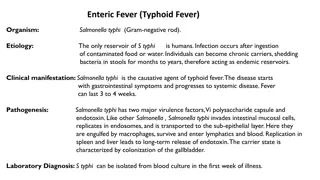
Salmonella are primary intestinal parasites. Causes : 1. Enteric fever (Typhoid fever by Salmonella typhi and paratyphoid fever caused by Salmonella paratyphi A, B and C ) 2. Gastroenteritis or food poisoning 3. Septicemia
EPIDEMIOLOGY Typhoid fever affects upto 30 million individuals world wide each year. Infection by S,typhi is more common in endemic areas where children & adolescents are most often affected. By contrast, S.paratyphi predominates in travelers & those living in developed countries.
PATHOGENESIS Incubation period : 7-10 days Infection by ingestion of contaminated food & water i.e feco-oral route Source of infection - case or carrier Convalescent person excrete organism in faeces & urine for 3-12 weeks Chronic carriers - organisms present in gall bladder & shed in faeces intermittently
MORPHOLOGY Affects primarily terminal ileum & colon GROSS: - Peyer's patches in terminal ileum become sharply delineated, plateau like elevations upto 8 cm in dia, with enlargement of draining L.nodes (mesenteric). - Peyer's patches involvement produces swelling, congestion & ulceration.
Shedding of mucosa & swollen lymphoid ts creates oval ulcer with their long axis along the length of the ileum. No significant fibrosis, hence fibrous stenosis seldom occurs. Main complication - perforation of the intestine & hemorrhage.
M/E Blunted villi Vascular congestion & edema Mononuclear inflammation - Macrophages( containing bacteria, RBC & nuclear debris), lymphocytes & plasma cells SPLEEN Gross- enlarged, soft, bulging M/E - Uniformly pale red pulp - Obliterated follicular markings - Prominent sinus histiocytosis and reticulo-endothelial proliferation
LIVER Typhoid nodule BONE MARROW & LYMPHNODE Typhoid nodule GALL BLADDER Typhoid cholecystitis which may be associated with gall stones chronic carrier state Also involves- kidneys (nephritis), bones (osteomyelitis), joints(arthritis), meninges (meningitis) & testis(orchitis)
CLINICAL FEATURES 1st week: fever, continuous type or step ladder type 2nd week: widespread mononuclear phagocyte invasion with rash (rose spots), abdominal pain & prostration. 3rd week: ulceration of peyer's patches, intestinal bleeding & perforation. Persistence of organism in gall bladder or urinary bladder passage of organism in urine or faeces CARRIER STATE ( source of infection to others)
LAB DIAGNOSIS A. Isolation and identification of causative agent from of patient blood, faeces, urine, bone marrow, bile, CSF - Bacteremia occurs early in the disease. Blood cultures are positive in 1st week in 90% cases, in 2nd week in 75% cases, in 3rd week in 60% cases & in 4th week and later in 25%cases
B. Demonstration of circulating antigens - by coagglutination test. C. Demonstration of antibodies in patient serum FELIX-WIDAL TEST Significant titers helps in diagnosis Serum agglutinins rise abruptly during the 2nd or 3rd week
TREATMENT Earlier Chloramphenicol was the drug of choice but produced plasmid mediated drug resistance and had side effects like aplastic anaemia Quinolones : Ciprofloxacin, Norfloxacin, Ofloxacin 3rd generation Cephalosporins : Cefotaxime, Cefoperazone, Ceftriaxone Cholecystectomy for carriers
LEPROSY (HANSEN DISEASE) It is a chronic infectious disease caused by Mycobacterium leprae, affecting mainly the skin & peripheral nerves & resulting in disabling deformities. M.leprae resembles M.tuberculosis but is less acid fast. Mainly involves the cooler parts of the body like skin, mouth, respiratory tract, peripheral nerves, superficial L.nodes & testis.
EPIDEMIOLOGY The disease is endemic in areas with hot & moist climates & in poor tropical countries. India accounts for one-third of all registered cases globally. In India, disease is seen more commonly seen in regions like TN, Bihar, Pondicherry, AP, Odisha, WB & Assam.
MODE OF TRANSMISSON Leprosy is a slowly developing communicable disease Incubation period between exposure to organism & appearance of signs varies from 2 to 20 yrs. Infectivity may be from following: - Direct contact with untreated patients - Materno-foetal transmisson - Infected mother milk to infant
CLASSIFICATION 1) LEPROMATOUS LEPROSY(LOW RESISTANCE ) 2) TUBERCULOID LEPROSY(HIGH RESISTANCE) 3) BORDERLINE LEPROSY
LEPROSY CURRENTLY CLASSIFED AS (Modified Ridley & Jopling s classification) TT-TUBERCULOID POLAR(HIGH RESISTANCE) BT BORDERLINE TUBERCULOID BB-MID BORDERLINE BL BORDERLINE LEPROMATOUS LL-LEPROMATOUS POLAR(LOW RESISTANCE)
Lepromatous leprosy (more severe form) Also known as anergic leprosy because of the unresponsiveness (anergy) of the host immune response. Cooler areas of skin are commonly affected. Skin lesions are generally symmetrical, multiple , hypopigmented & erythematous macules, papules, nodules or diffuse infiltrates. Nodular lesions may coalesce to give leonine facies. Lesions are hypoaesthetic/anaesthetic but sensory disturbance is not that distinct.
TUBERCULOID LEPROSY Skin lesions occur as single/few asymmertical, hypopigmented, erythematous macules. Sensory disturbance is distinct.
LEPROMATOUS LEPROSY In the dermis there is proliferation of lipid laden macrophages particularly around bld vessels, nerves & dermal appendages. The foamy marophages are known as lepra cells or Virchow cells. Lepra cells are heavily laden with acid fast bacilli (globi or cigarettes - in - pack) Dermal infiltration of lepra cells separated from epidermis by subepidermal uninvolved clear zone. Epidermis overlying the lesion is thinned out , flat or may even ulcerate.
TUBERCULOID LEPROSY Dermal lesions show granulomas resembling hard tubercles composed of epithelioid cells , langhan s giant cells & a peripheral rim of lymphocytes. Granulomatous infiltrate erodes the basal layer of epidermis ie there is no clear zone. Lepra bacilli are few Lesions have predilection for dermal nerves which may be destroyed.
LL TT SKIN LESIONS Symmetrical,multiple, hypopigmented, erythematous, maculopapular or nodular (leonine facies) Present but sensory disturbance is less severe Asymmetrical, single or a few lesions, hypopigmented & erythematous macules Present with distinct sensory disturbance NERVE INV H/P Collection of foamy macrophages or lepra cells in the dermis separated from epidermis by a clear zone Hard tubercle eroding the basal layer of epidermis, no clear zone
BACTERIOLOGY Lepra cells are highly positive for lepra bacilli seen as globi or cigarettes- in pack appearance Lepra bacilli few, seen in destroyed nerves as granular or beaded forms IMMUNITY Suppressed (low resistance) Good immune response (high resistance) LEPROMIN TEST Negative Positive
DIAGNOSIS OF LEPROSY 1) Diagnosis of leprosy is most commonly based on the clinical signs and symptoms. In an endemic country or area, an individual should be regarded as having leprosy if he or she shows ONE of the following cardinal signs: skin lesion consistent with leprosy and with definite sensory loss, with or without thickened nerves positive skin smears
2) For patients presenting the above symptoms, a leprosy diagnosis is confirmed after analysis of tissues biopsied from infected sites. Histopathological diagnosis is made Typically, a stain for acid-fast bacteria is performed . Additional steps may include - Culturing infected tissue to confirm other bacteria are not involved - M. leprae cannot be cultured in vitro -Using PCR
3) OTHER TESTS Lepromin skin test Polymerase chain reaction (PCR)
Frequently asked Questions a) Difference between Lepromatous leprosy & Tuberculoid leprosy 37
Reading List/Reference List Robbins Basic Pathology: Kumar, Abbas, Aster, 9th Edition Essential Pathology for Dental Students: Harsh Mohan, Sugandha Mohan, 5th Edition 38


")


")


 For patients presenting the above symptoms,")
 For patients presenting the above symptoms,")
 OTHER TESTS")