Understanding Chromosomes: Structure, Function, and Inheritance
Explore the essential aspects of chromosomes, including their structure, functions, types, and role in determining gender and genetic disorders. Learn about chromatin, single and duplicated chromosomes, chromatids, centromeres, and autosomal abnormalities. Understand how chromosomes determine the gender of offspring and the importance of the correct number of chromosomes in reproductive cells for proper development.

Download Presentation

Please find below an Image/Link to download the presentation.
The content on the website is provided AS IS for your information and personal use only. It may not be sold, licensed, or shared on other websites without obtaining consent from the author. If you encounter any issues during the download, it is possible that the publisher has removed the file from their server.
You are allowed to download the files provided on this website for personal or commercial use, subject to the condition that they are used lawfully. All files are the property of their respective owners.
The content on the website is provided AS IS for your information and personal use only. It may not be sold, licensed, or shared on other websites without obtaining consent from the author.
E N D
Presentation Transcript
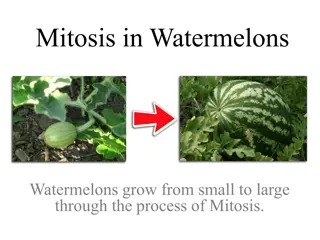
UNIT 2 THE CHROMOSOMES AND KARYOTYPE PHASES OF MITOSIS AND MEIOSIS
GLOSSARY Chromatin - diffuse, long fibers of DNA with proteins attached Single chromosomes - a chromosome which has not been duplicated (like of an x) Duplicated chromosomes - a chromosome which has been duplicated (a complete X) Chromatid - one half of a duplicated chromosome, looks like a single chromosome Centromere - a structure made of protein which holds the two halves of a duplicated chromosome together.
CHARACTERISTICS OF CHROMOSOMES 1. Location and Function Located in the center of cells called the nucleus Each chromosomes features protein and a single DNA molecule. The DNA wrapped around histones (spool-like proteins) A key part of the process of accurately copying DNA and distributing in many cell divisions
CHARACTERISTICS OF CHROMOSOMES 2. PAIRS (23) Male 46 chromosomes 22 pairs of chromosomes Different pattern of sex chromosomes (XY) One of each pair autosomes and an X is maternal origin The father contributes the Y and the remaining autosomes Female 46 chromosomes Two X chromosomes One of each pair of the autosomes and one X is of maternal origin The other 23 are of paternal origin
CHARACTERISTICS OF CHROMOSOMES 3. Child Gender The mother always contributes an X chromosomes to her child The father of the contributes either an X chromosomes or a Y chromosomes As a result, the father is the parent who determines the sex of a child. Still, a child inherits some traits from his mother and other traits from his father.
CHARACTERISTICS OF CHROMOSOMES 4. Autosomal Types 1 to 22 = autosomal chromosomes Reproductive cells such as eggs and sperm must have the right number of chromosomes in order to have offspring that develops correctly. Example: Down Syndrome 3 copies of chromosome 21 instead of two copies Autosomal abnormality
KARYOTYPES KARYOTYPES DEFINITION Refers to a picture of individual s chromosomes. USES 1.Use to detect fetal abnormalities 2.Form of genetic screening conducted on potential parents. KARYOTYPING A technique that uses a picture of an individual s chromosomes to analyze and detect chromosomal abnormalities
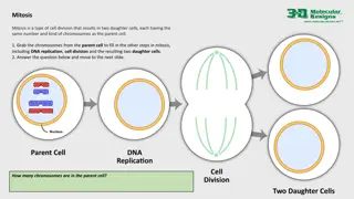
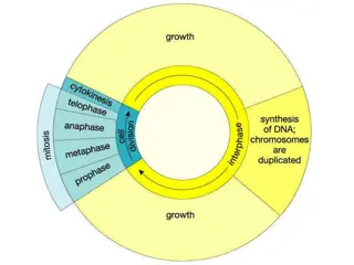
WHAT IS MITOSIS? is the process, in the cell cycle, by which a cell duplicates into two genetically alike daughter cells. In mitosis, chromosomes in the cell nucleus are separated into two identical sets of chromosomes, each in its own nucleus. Vital for tissue formation and maintenance
PHASES/STEPS OF MITOSIS 1. Prophase 2. Metaphase 3. Anaphase 4. Telophase
PROPHASE The first stage of mitosis Chromatin condenses to form duplicated chromosomes The nuclear envelope breaks down (degenerate) The spindle apparatus (made by microtubules) forms The spindle attaches to the centromeres of the chromosomes and moves the chromosomes around during cell division
METAPHASE The second stage The mitotic spindle moves the duplicated chromosomes so that they are lined up at the cell equator (middle of the cell) equidistant from the two poles (opposite ends) of the cell.
ANAPHASE The third stage Centromeres of duplicated chromosomes divide and split The mitotic spindle pulls the sister chromatids separate from each other as they move to the opposite poles of the cell
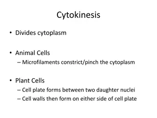
TELOPHASE The fourth stage Duplicated chromosomes decondense back into chromatin The nuclear envelope begins to form around the chromosomes so that the two separate daughter nuclei are created The mitotic spindle breaks down Two daughter cells are produced Diploid cells (2N)- 46 chromosomes same as the mother cells
MITOSIS AND CANCER DNA, sometimes called a genetic blueprint, contains the hereditary material in nearly all organisms. Two types of errors, or mutations, can occur when DNA is improperly copied: silent mutations, which have no impact on the DNA sequence, and "missense" mutations, which change a DNA sequence and often impact the associated function. Missense mutations can multiply over time, leading to cell cycle disruption and formation of tumors -- cells that don't stop dividing. Cancer occurs when the normal "checkpoints" regulating mitosis are ignored or overriden by a cancer cell, resulting in uncontrolled cell division.
WHAT IS MEIOSIS? Types of cell division that creates egg and sperm cells Ensures that humans have the same number of chromosomes in each generation. A two step process that reduces the chromosomes number by half 46- 23 (to form sperm and egg cells)
Meiosis 1: prophase 1, metaphase 1, anaphase 1, and telophase 1 Meiosis 2: prophase 2, metaphase 2, anaphase 2, and telophase 2
In the first meiotic division, the number of cells is doubled but the number of chromosomes is not. This results in 1/2 as many chromosomes per cell. The second meiotic division is like mitosis; the number of chromosomes does not get reduced.
PROPHASE I OF MEIOSIS I CROSSING OVER the DNA coils tightly and individual chromosomes becomes visible via microscope Homologous chromosomes become closely associated in synapsis and they exchange segments by crossing over
METAPHASE 1 OF MEIOSIS 1 The disappeared chromosomes equatorial plane. nuclear membrane and move has the to
ANAPHASE 1 OF MEIOSIS 1 Two members off each bivalent disjoin, one going to each pole These bivalent are assorted independently to each pole The cytoplasm divides Each cell now has 23 chromosomes Each of which has a pair of chromatids differing from one another as a result from crossing over
TELOPHASE I OF MEIOSIS I The nuclear membrane reforms around the daughter nuclei Each daughter nucleus contain two sister chromatids for each chromosomes, attached to a common centromere Because of crossing over, the two sister chromatid are not identical.
PROPHASE 2 OF MEIOSIS 2 The nuclear envelope breaks down and a new spindle fiber forms
METAPHASE 2 OF MEIOSIS 2 Spindle fibers bind to both sides of centromere
ANAPHASE 2 OF MEIOSIS 2 The spindle fibers contract and the sister chromatid move toward opposite poles.
TELOPHASE 2 OF MEIOSIS 2 Nuclear envelopes reform around the sets of daughter chromosomes
MEIOSIS THUS HAS 3 IMPORTANT CONSEQUENCES 1.Gametes contain only one representative of each homologous pair of chromosomes. 2.There is a random assortment of paternal and maternal homologous. 3.Crossing- over ensures uniqueness by further increasing genetic variation.
DISORDERS DUE TO MEIOSIS Errors due during segregation (chromosomes segregates too many or few chromosomes in egg or sperm) trisomy = an extra chromosome of a particular pair in each cell monosomy = one chromosome fewer in each cell For example Trisomy 21 Down syndrome cells have an extra copy of chromosome 21 (trisomy 21). Turner syndrome cells have only one X chromosome (monosomy X) and no Y chromosome


")